inForm® Tissue Finder™
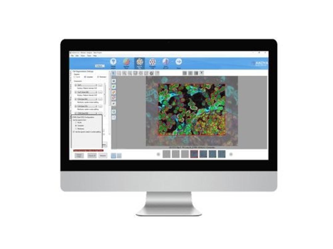
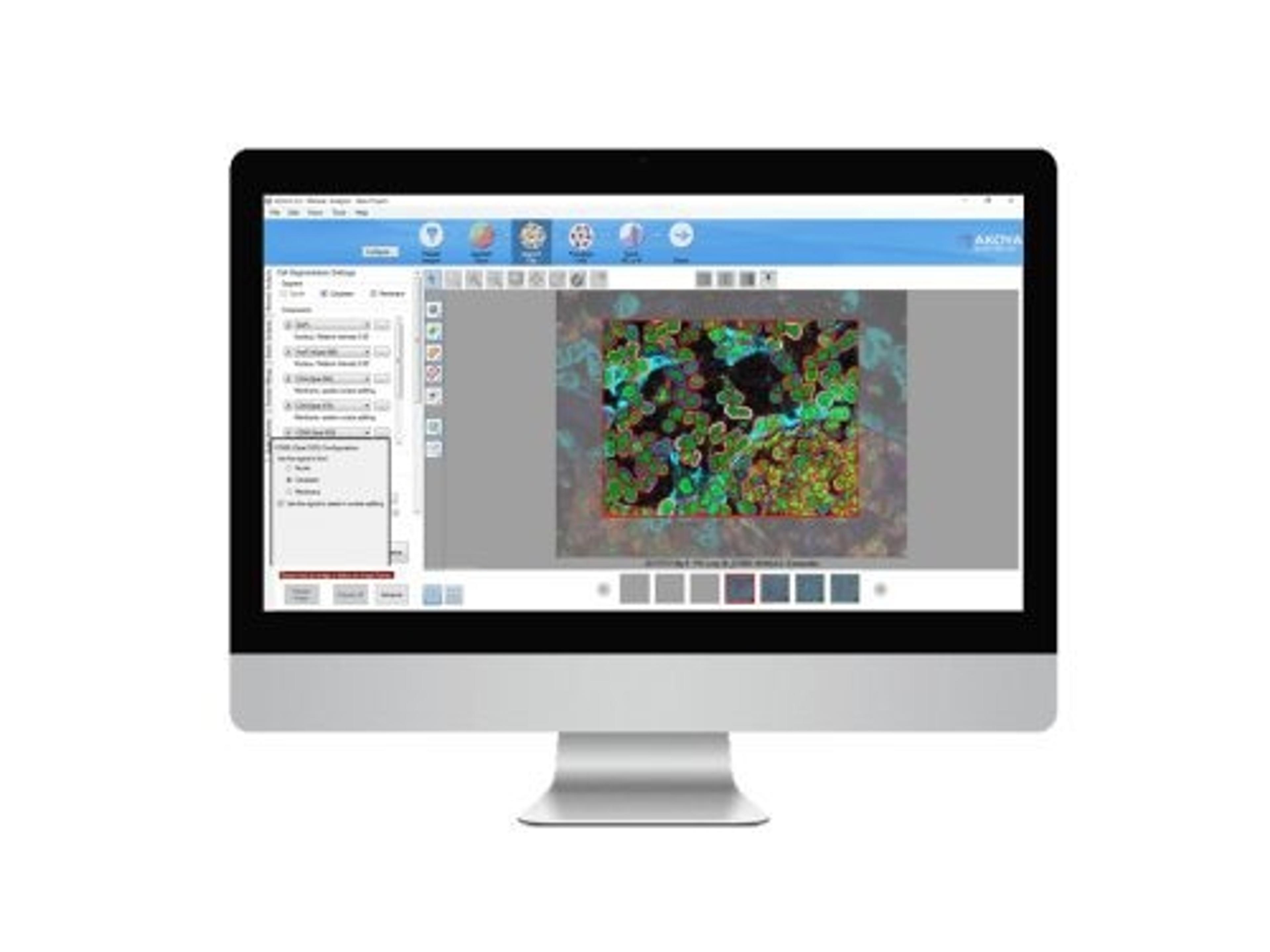
inForm® Tissue Finder™ adds exceptional functionality to inForm Cell Analysis to automate the detection and segmentation of specific tissues through powerful patented pattern recognition algorithms. Automation provides consistent reproducible results and enables comparative studies of multiple markers and specimens, supporting researchers to make faster discoveries of the indicators of disease.

The supplier does not provide quotations for this product through SelectScience. You can search for similar products in our Product Directory.
A lifesaver for vectra analysis.
Unmix mIHC (vectra) images and downstream signal scoring and phenotyping analysis
InForm is the best platform for mIHC image unmixing. The software is trainable, intuitive and easy to use. The batch analysis function provides opportunities to standardize analysis parameters across multiple cohorts. Highly reliable and reproducible results.
Review Date: 3 Dec 2020 | Akoya Biosciences
Once trained, inForm will locate and analyze user-specified regions automatically across an entire image or multiple images. Large numbers of images can be rapidly batch processed, allowing analysis that might have taken days to be done in a matter of minutes. Visualizes, analyzes, quantifies and phenotypes immune and other cells in situ in solid tissue Pathology Views™ to render immunofluorescence images as H&E, DAB and hematoxylin Automated quantitation of biomarker expression from specific tissue types (e.g. tumor/stroma) Trainable tissue segmentation learns by example – automatically locates tissues / structures of interest Separates weakly expressing and overlapping markers Batch processing of images using customizable image analysis workflows Scoring (% positivity, 0/1+/2+/3+, co-localization and more) Review and merge data from a set of images or slides into summary data files to assure data quality Export data and images for further analysis e.g. TIBCO Spotfire ® Analyst for Quantitative Pathology Supports Akoya's Phenoptics™ 2.0 workflow solution for cancer immunology research.

