Lux3 BR brightfield live cell imager
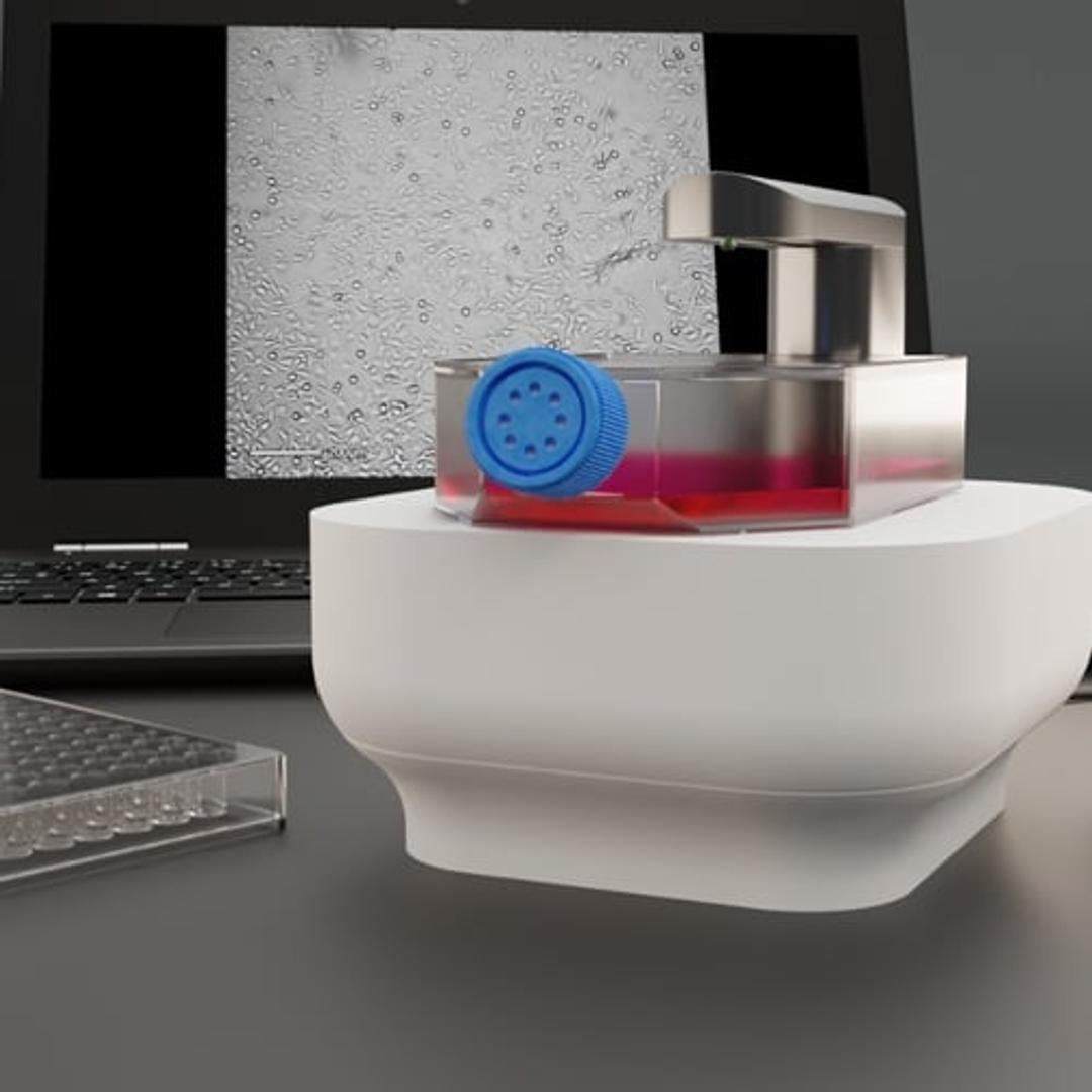
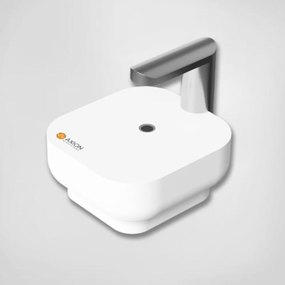
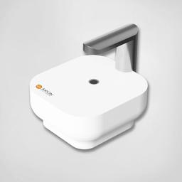
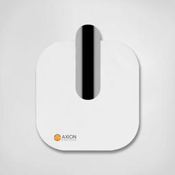
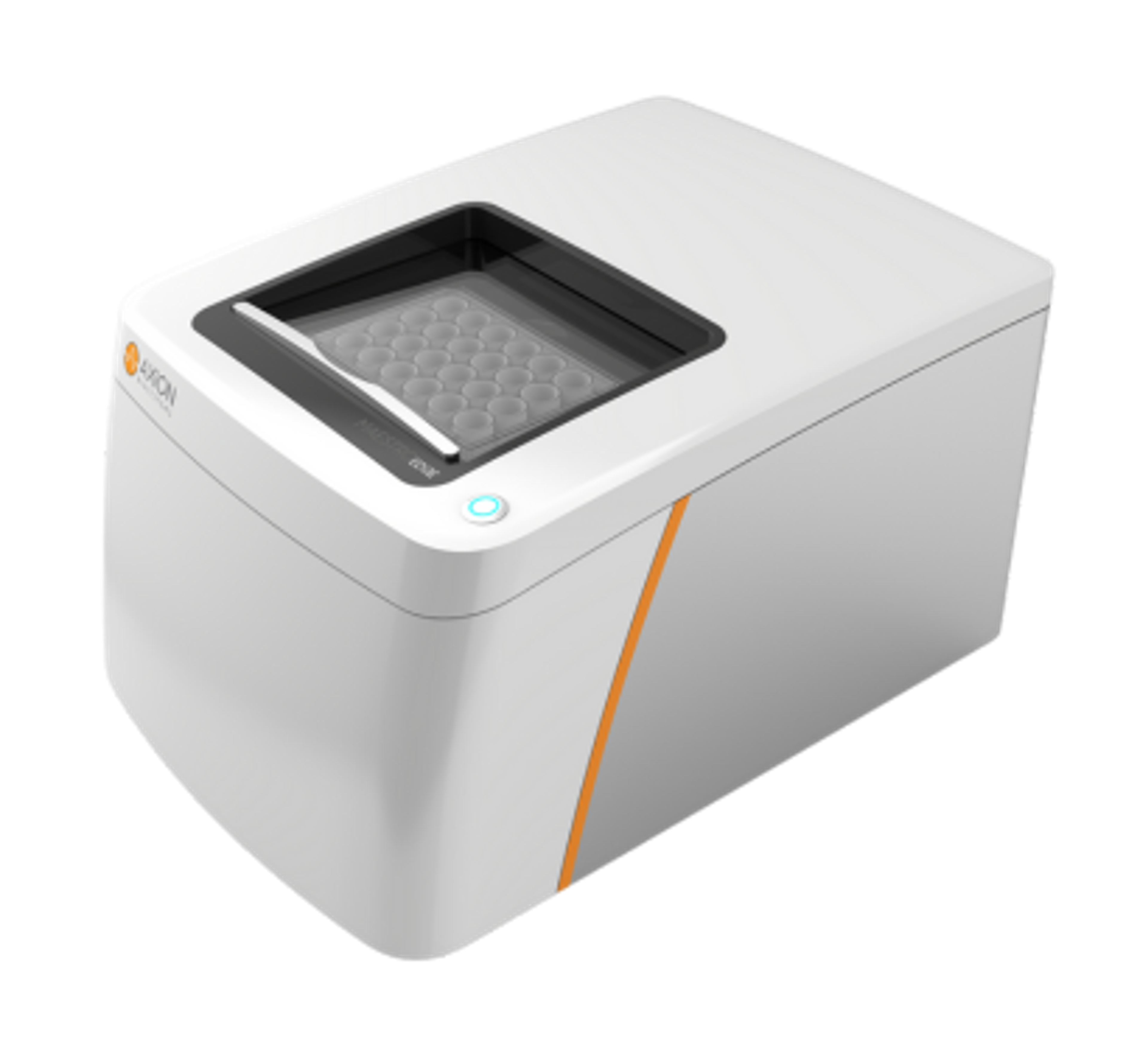
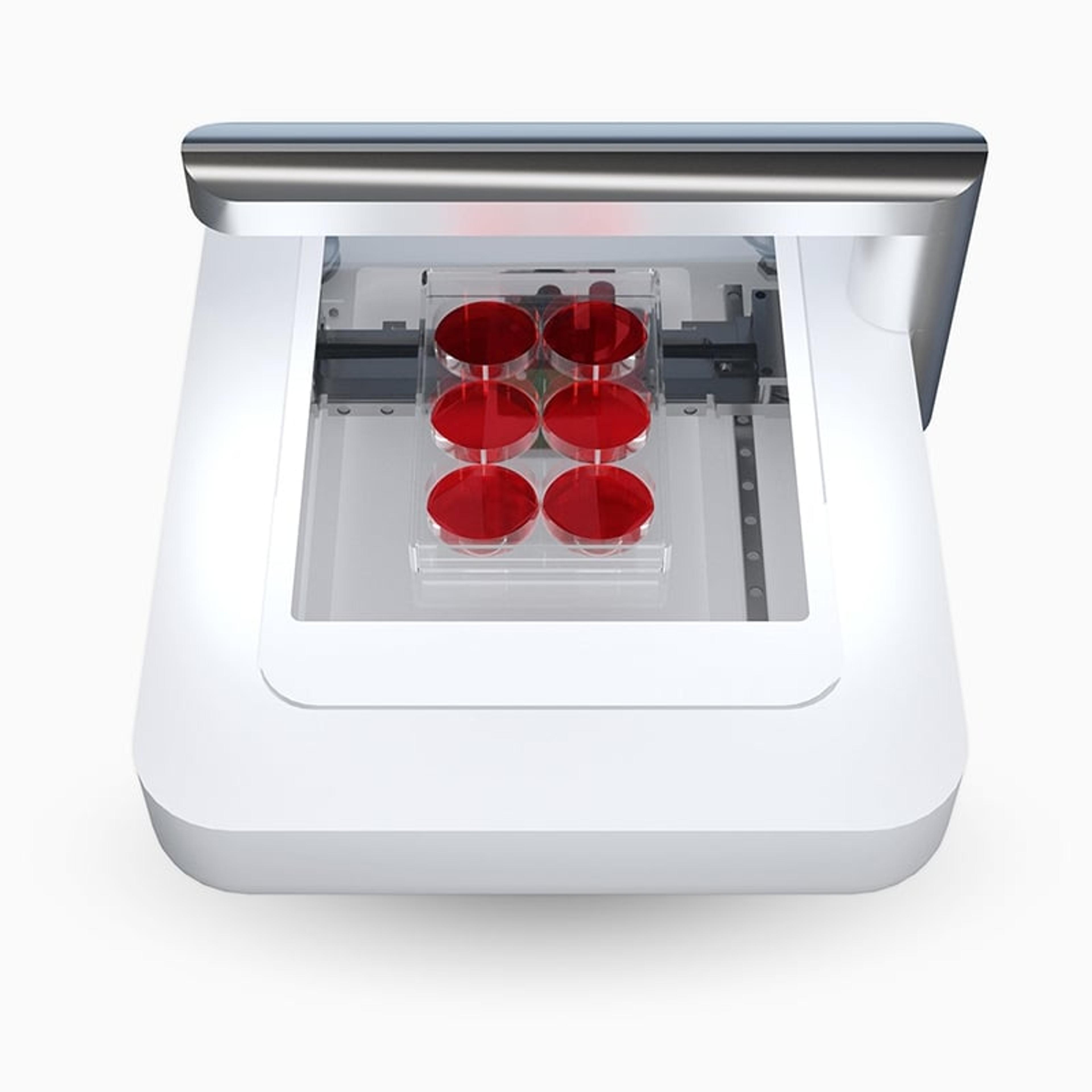
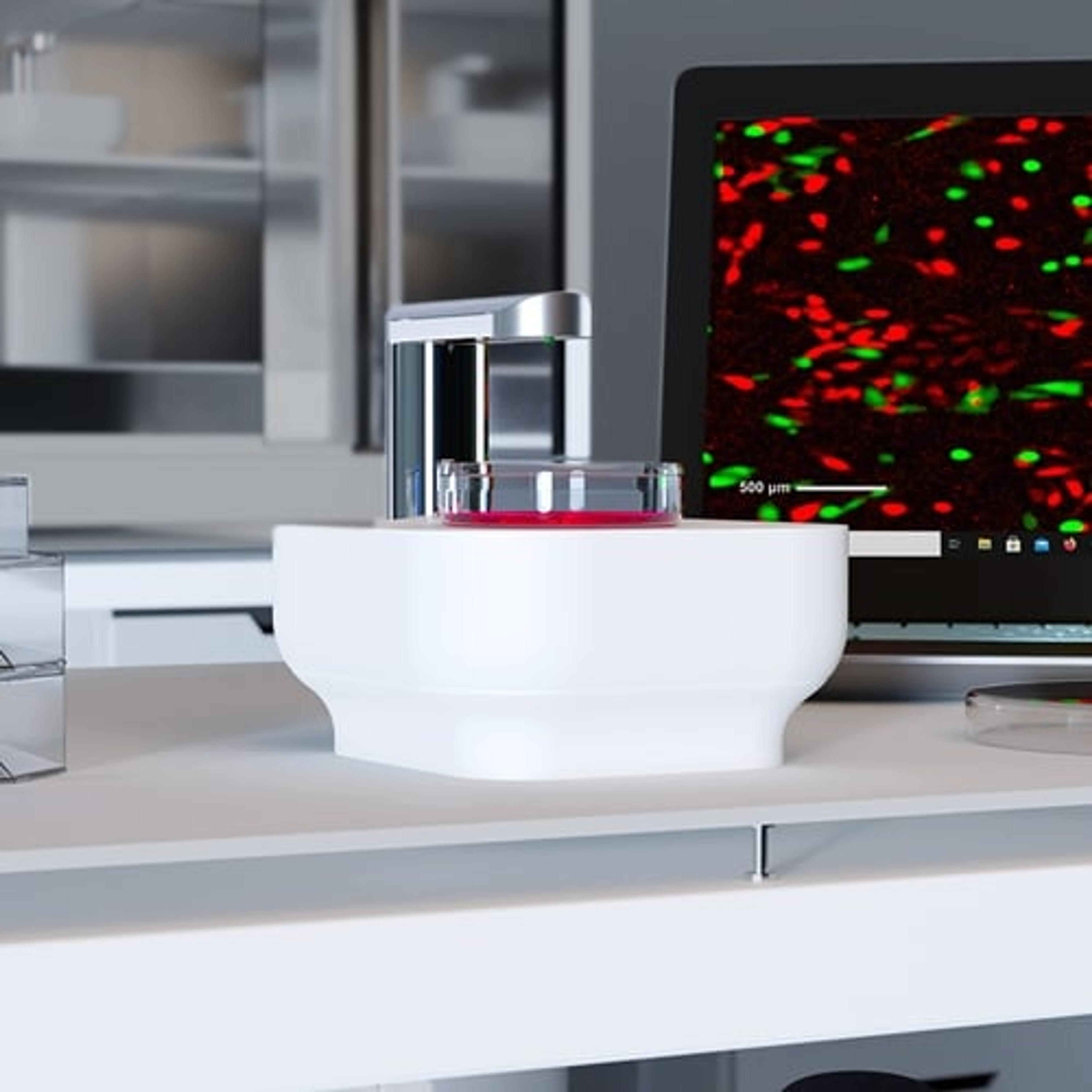
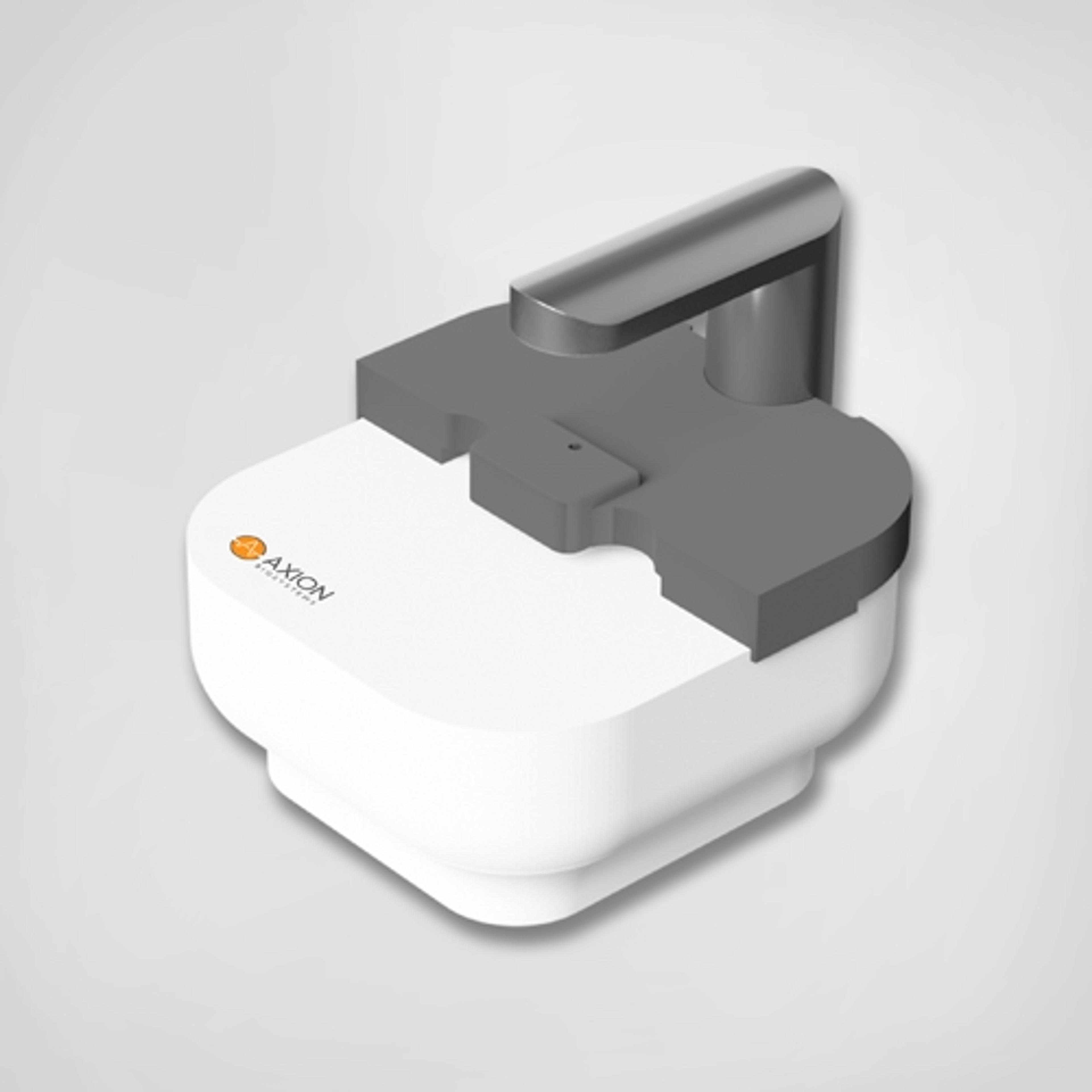
Lux3 BR is a small brightfield live-cell imager, equipped with a high-quality 6.4 MP CMOS camera. It is designed to work inside a standard cell culture incubator without disturbing temperature, airflow, and optimum culture conditions. Detailed brightfield images can be captured using the new device. In both x- and y-direction, 2072 pixels combined with 1.45 mm field of view provide a resolution of 0.7 µm/pixel.
Efficient device and offers very good results
The effect of anthocyanin-rich fruits on fibroblasts
The device is very efficient and easy to use. Its benefits are that the classic methods of determining cell viability (MTT, MTS, Trypan blue) can be replaced and thus save time and keep samples/cells intact. Another advantage is the software of the device that can measure the surface of the wound, and the closure of the wound and thus replaces the sophisticated mathematical calculation. With the help of CytoSMART Lux 2, cell growth and the confluence of fibroblasts can be recorded and at the same time, cell morphological changes can be observed under different treatments.
Review Date: 18 Jun 2022 | Axion BioSystems
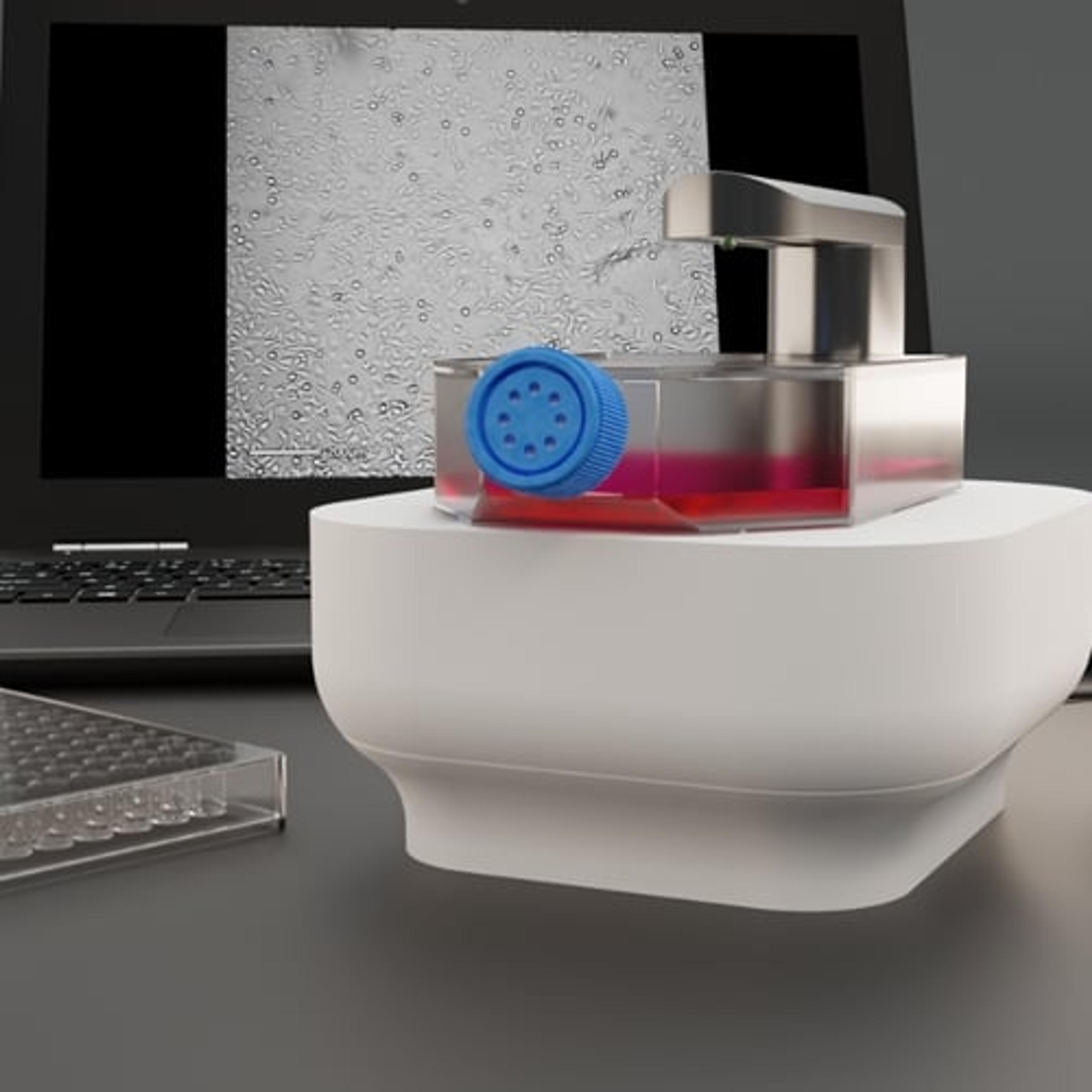
Lux3 BR is a small brightfield microscope, equipped with a high-quality 6.4 MP CMOS camera. Unlike the high-end microscopes with incubation box, the Lux3 BR ensures that cultures are not exposed to temperature, humidity, or CO2 fluctuations that can stress the cells, as the device easily fits in every CO2-incubator.
The Lux3 BR is ideal for capturing crisp brightfield images and videos of living cells. The image size of 2072×2072 pixels combined with the 1.45×1.45 mm field of view provide a resolution of 0.7 µm/pixel. Even at the commonly required image resolution of 300 dpi for printed scientific publications, these images can fill the entire page width if desired, without compromising the image quality.
The main features and benefits of the Lux3 BR include:
- High-quality images and time-lapse movies to investigate the development of cellular processes
- Incubator- and laboratory-friendly device due to its compact size and open design
- Remote data access via the Cloud with a smartphone, tablet, or laptop outside the lab
- Flexibility to expand a single live-cell imaging system to Duo and Multi Kits
Images and videos of running or completed experiments can be accessed, processed, and analyzed from any desired location using the Cloud-based environment. Therefore, cells can be monitored without having to open the incubator, or even be in the lab. The integrated cloud-based image analysis facilitates quantification of output parameters, e.g., cell confluence or wound healing rate. The automated quantification minimizes avoidable variation and bias in the interpretation of results.
A setup with the Lux3 BR can be easily expanded to two or even four devices that can be operated and controlled individually via a single laptop. Since both Lux3 BR Duo Kit and Multi Lux3 BR devices can be placed directly next to each other in the same incubator, it allows for systematic comparison of control and treated samples, as all monitored cell cultures are maintained in an identical culture environment.

