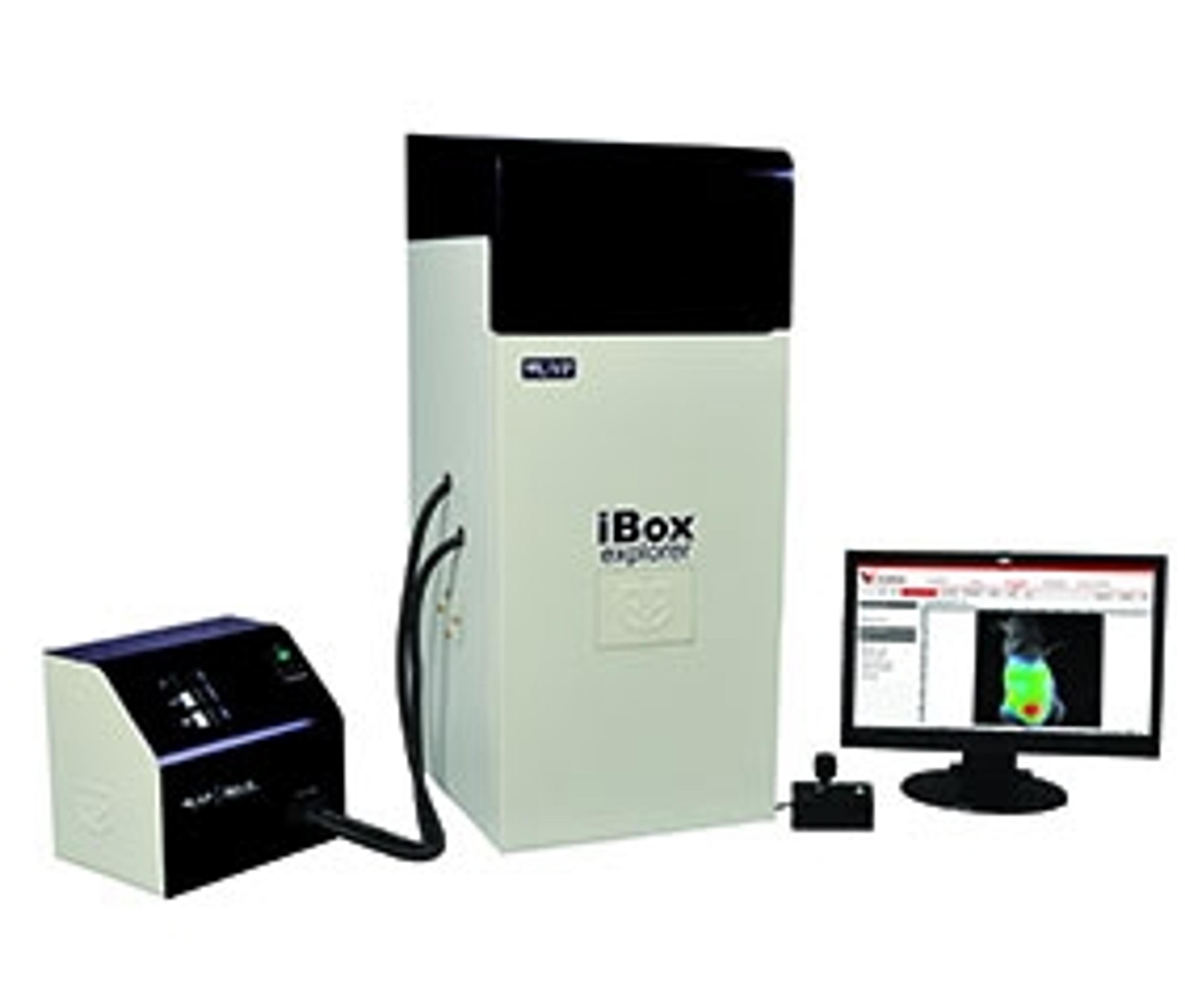
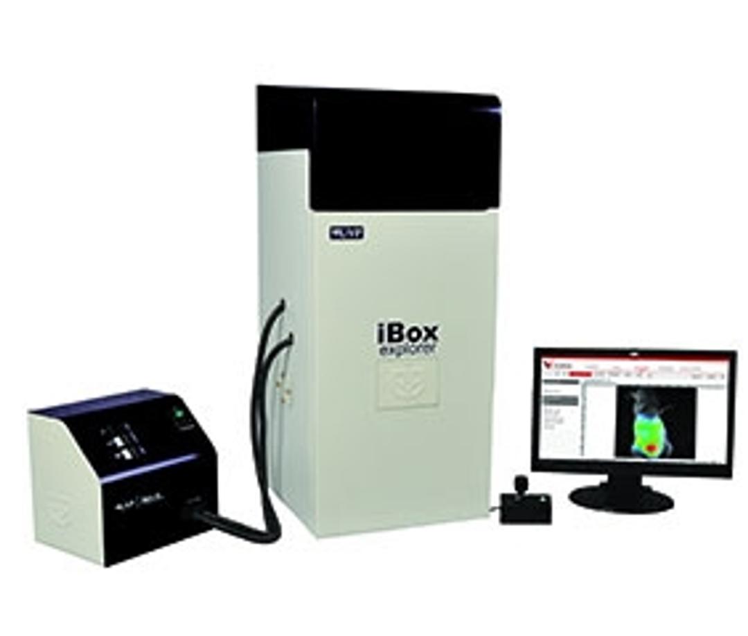
UVP iBox Explorer™ 2 Imaging Microscope
Macro to Micro Fluorescent In Vivo Imaging - Now researchers can take detection of fluorescent markers in small animals to a new level with the UVP iBox Explorer 2 Imaging Microscope!

Receive your quote directly from the manufacturer.
The results are really reliable with great accuracy.
Analysis of microorganisms like microbacterium tuberculosis in the laboratory
The product is quite easy to use and does not need comprehensive understanding.
Review Date: 6 Jun 2019 | UVP, An Analytik Jena Company
I am getting very crisp images.
Microbiome research
Excellent machine with multiple features. It can count bacteria, take gel images and it is very good for fluorescent imaging. It is very convenient to use and doesn't have complex software.
Review Date: 23 Aug 2017 | UVP, An Analytik Jena Company
The UVP iBox Explorer 2 is unique in its ability to view macro to micro in the whole animal to an individual cell, subcutaneously, and within the body cavity of mice. The upright optics provide an ultra-long working distance and high numerical aperture (NA) for detailed fluorescent in vivo imaging. UVP iBox Explorer² is an ideal system for imaging the distribution of fluorescent markers within small animals for pre-clinical research. Imaging of the whole animals is possible and can accommodate up to two mice. The cooled, scientific-grade CCD camera rapidly captures publication-quality images. A bright xenon arc lamp provides excitation light from the visible to NIR range, accommodating a host of in vivo studies and fluorescent markers. Application-specific filter sets can be tailored for imaging in pre-clinical fluorescence studies. Features: Magnification ranges of 0.17x - 16.5x enables an easy transition from the macroscopic to the microscope scale. UVP iBox Explorer 2 enables researchers to visualize micro-injection of cancer cells in vivo. Ability to image organs and cells subcutaneously and within the body cavity of living mice. Optical configurations are parcentered and parfocal, allowing seamless imaging through the magnification ranges. Leading-edge high frame rate cooled color camera enables quick detection, image capture and high throughput. Easy-to-use software automates research with templates for reproducible, consistent results. Bright illumination of samples produces an intense fluorescent signal and fast exposure times. An unlimited selection of filters enables users to image in the fluorescent, visible, and NIR ranges for multiple applications (RFP/GFP included, others optional). Generate accurate data with VisionWorks® Software.