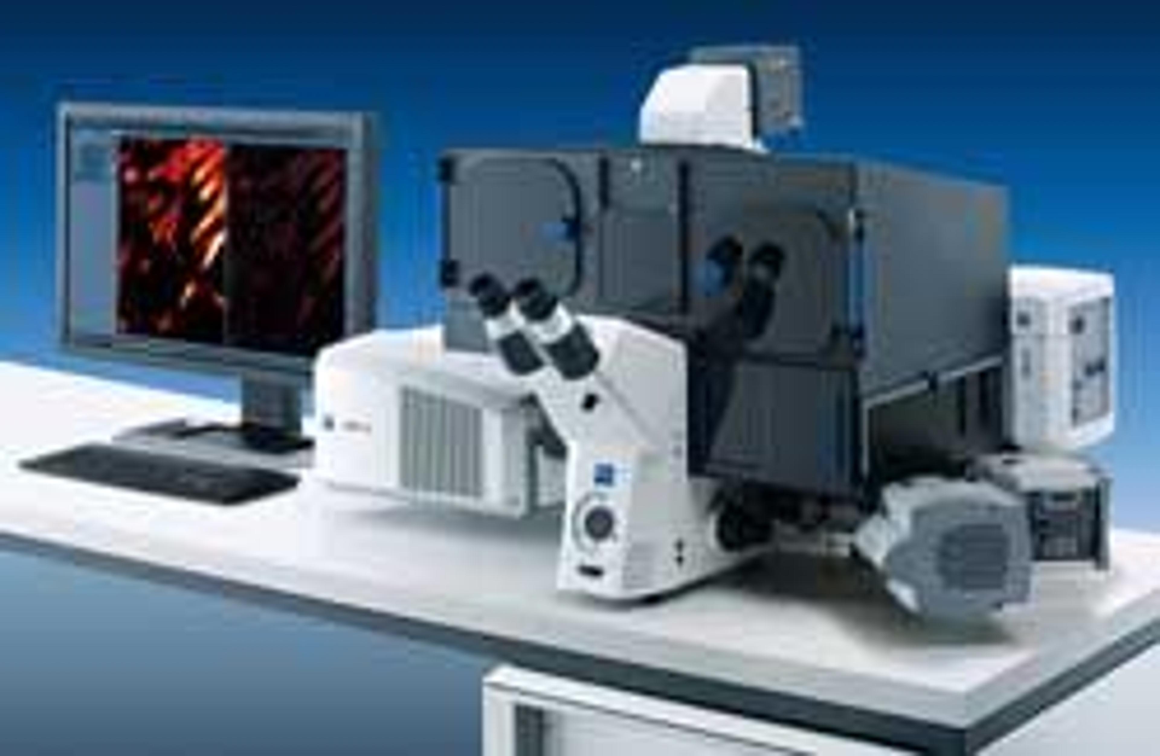
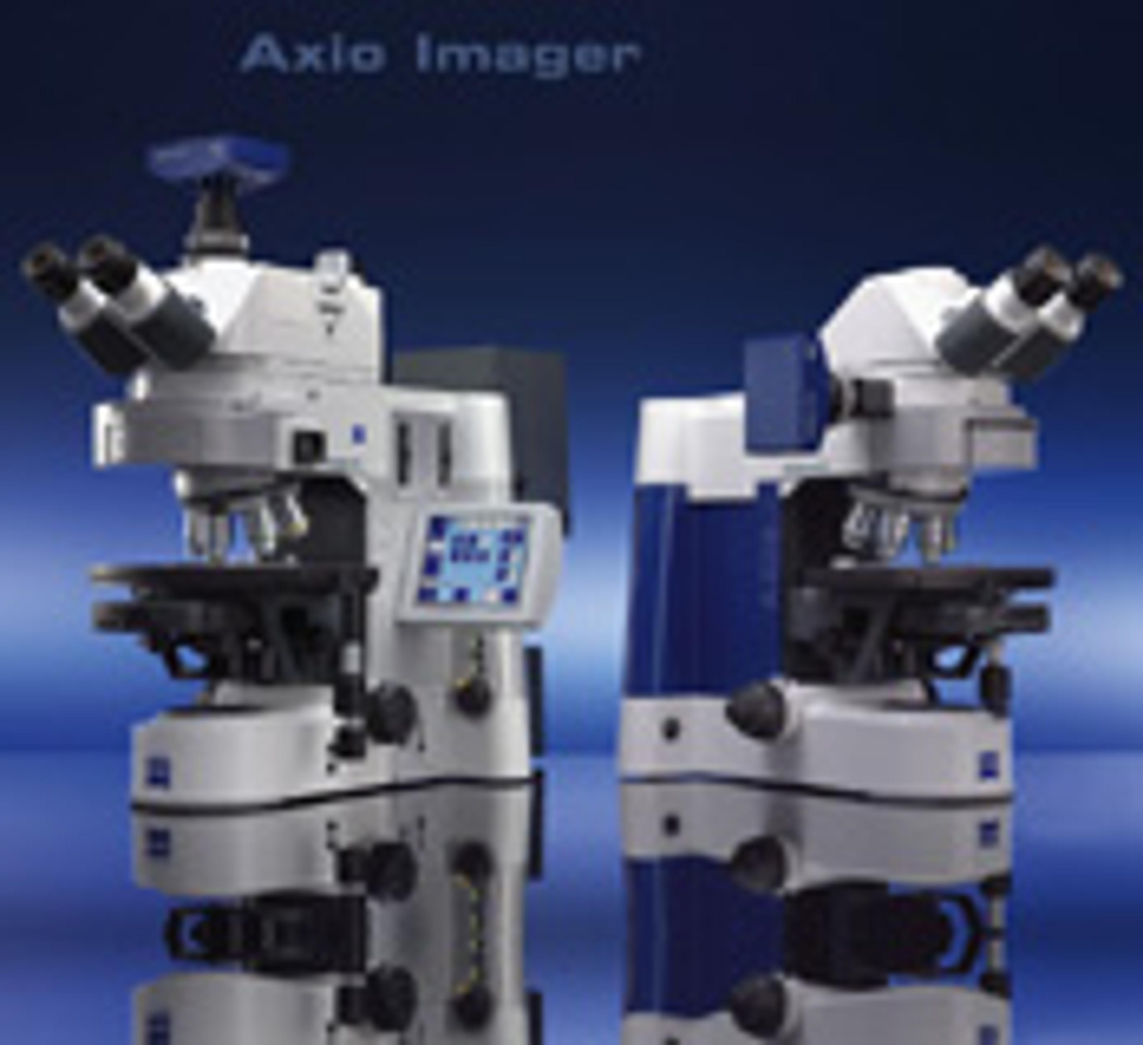
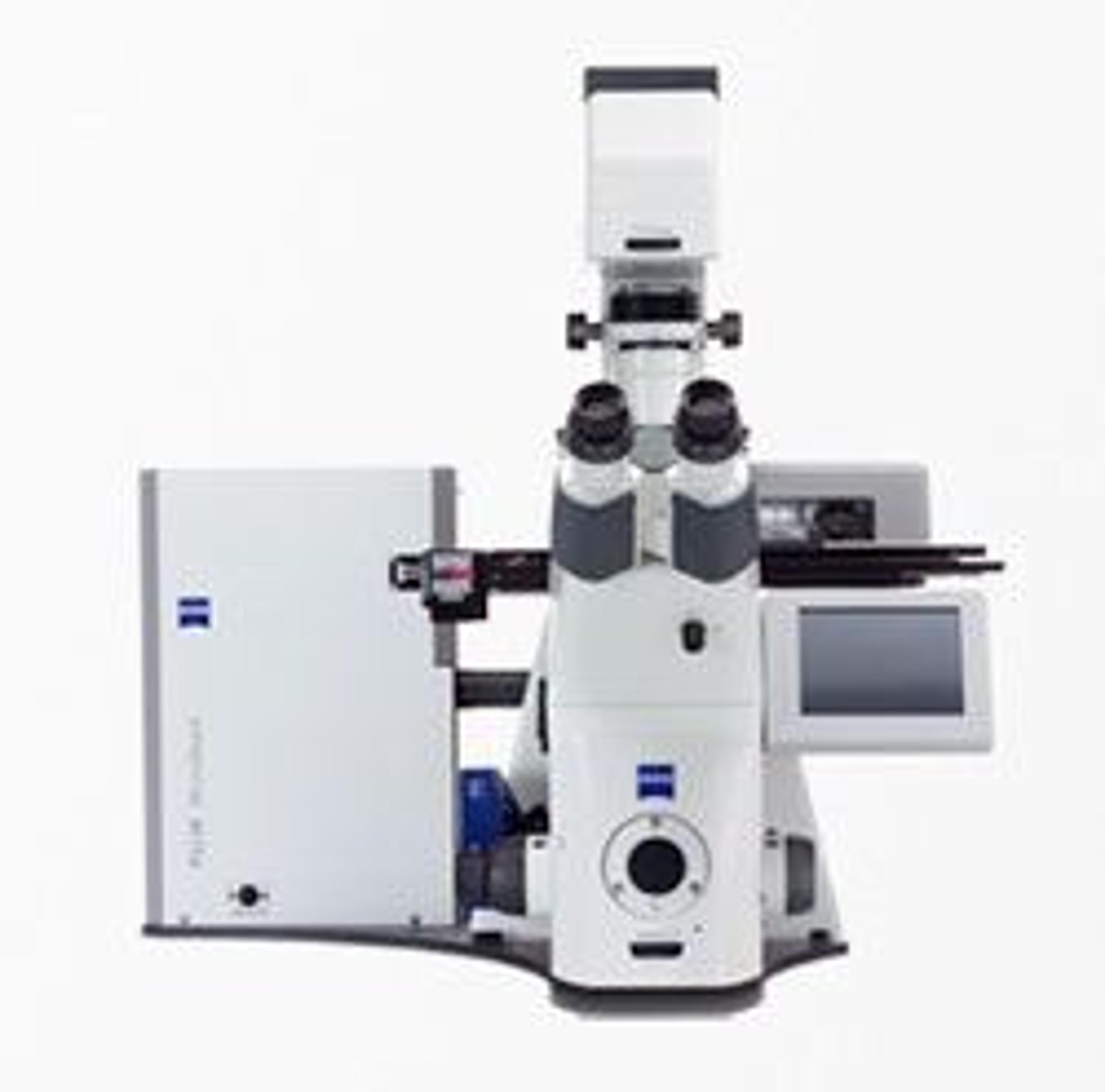
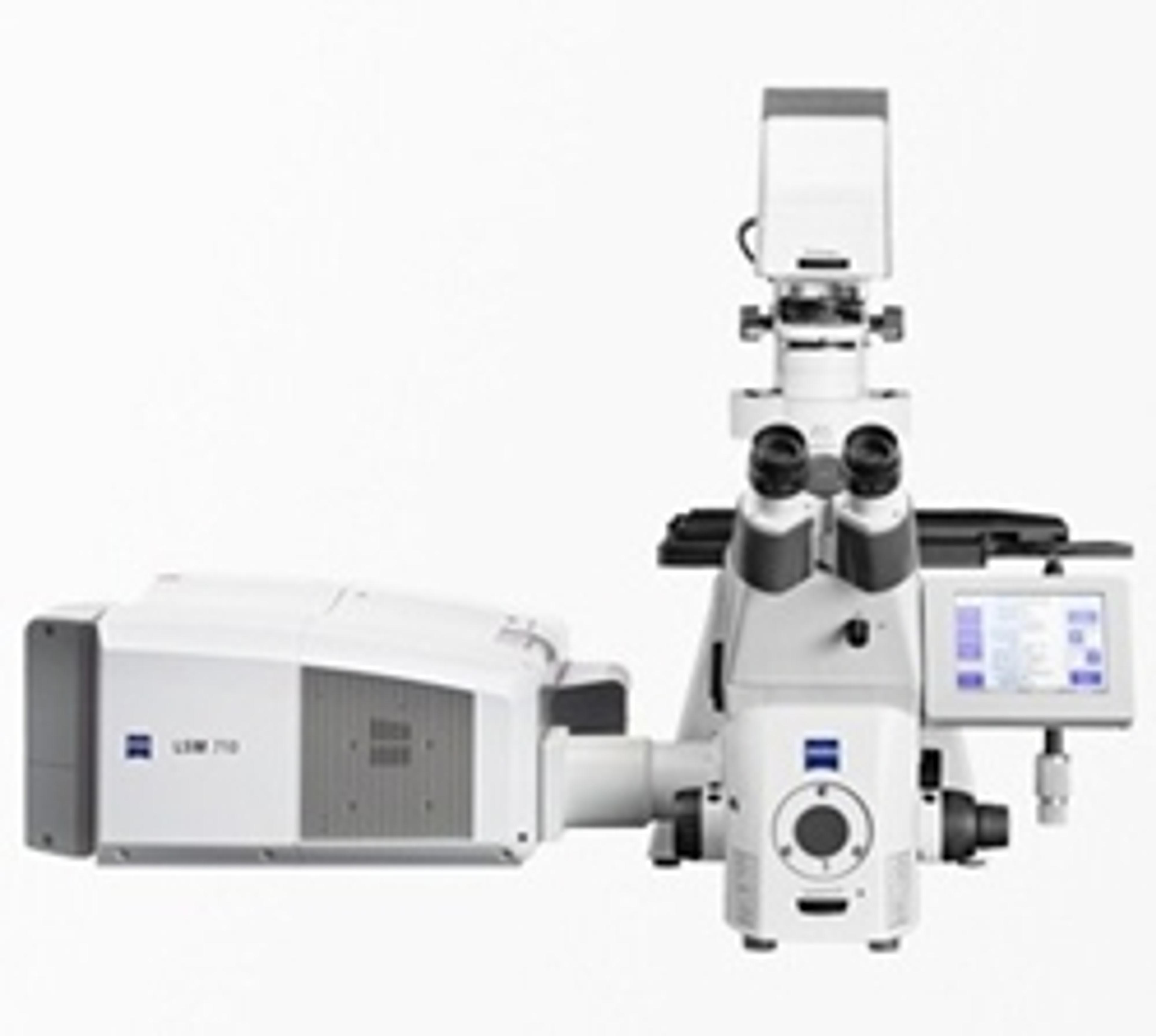
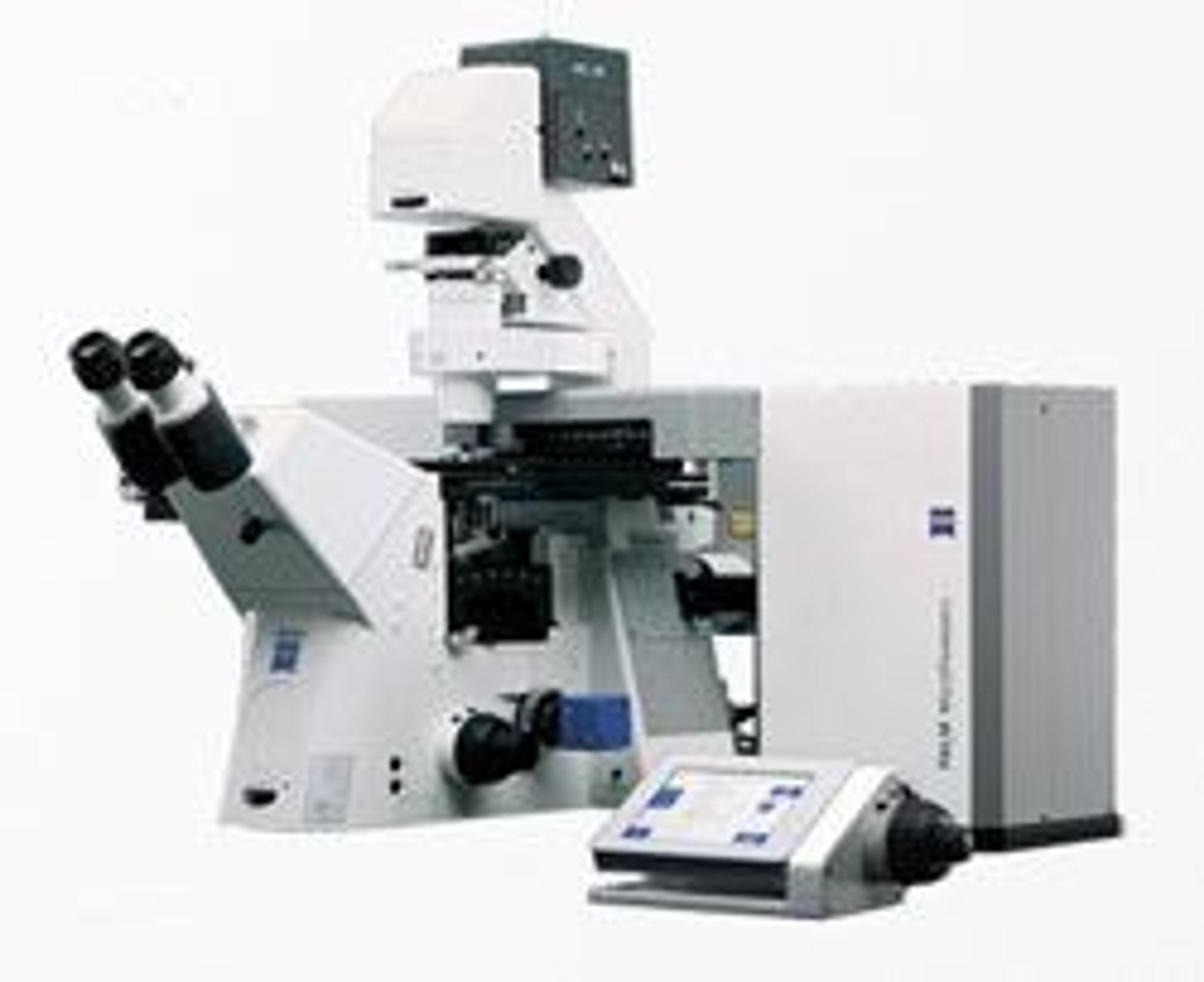
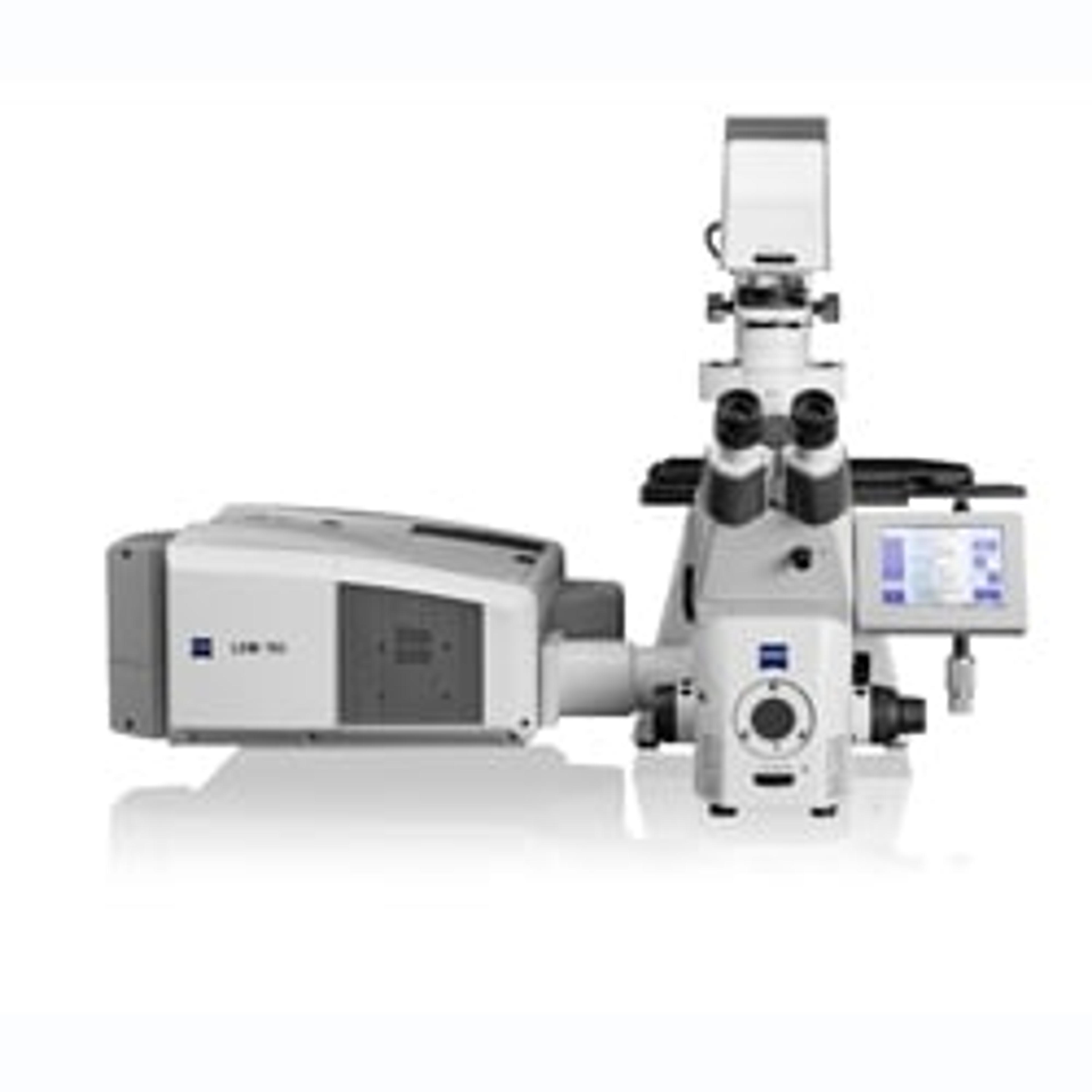
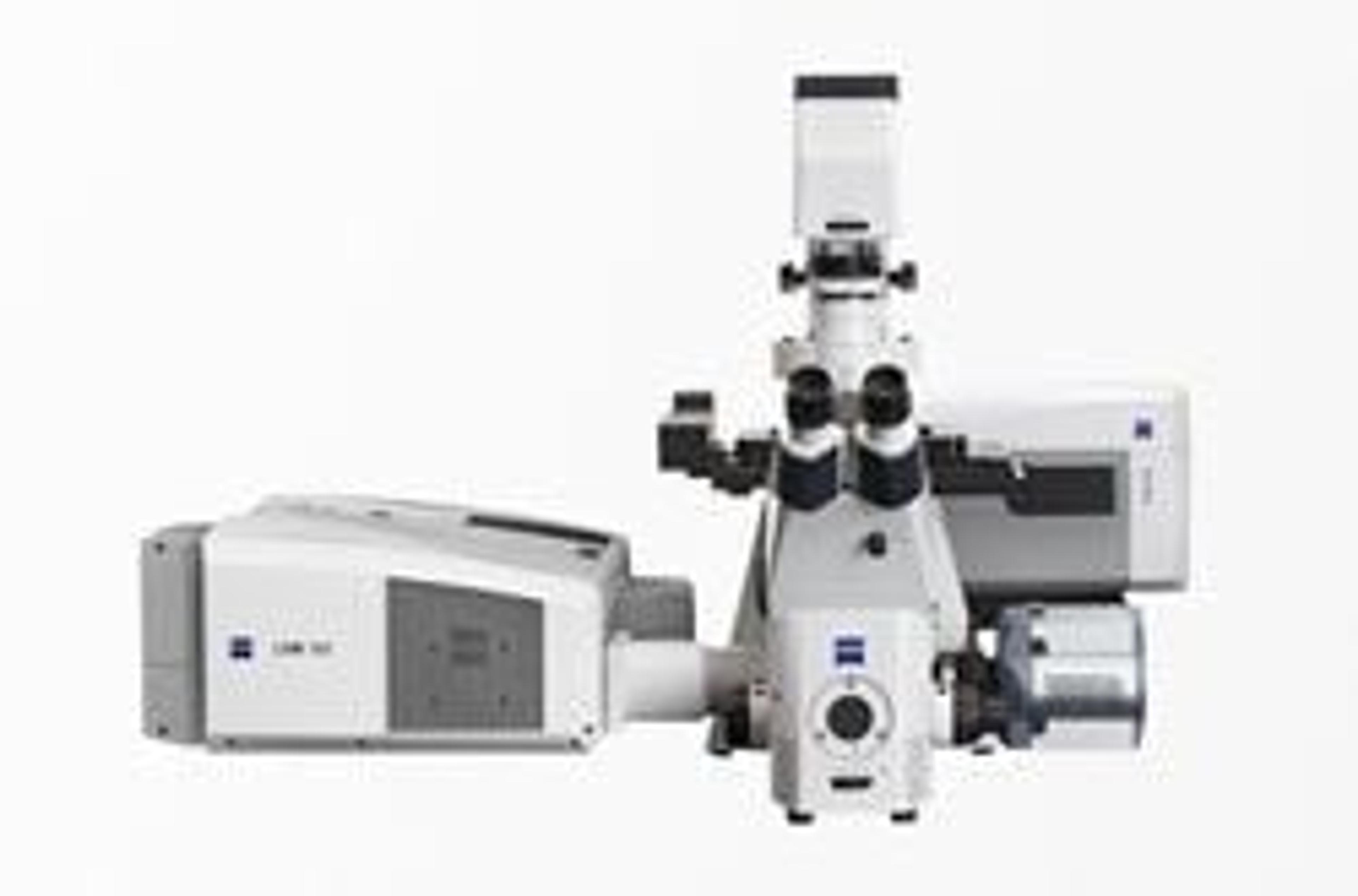
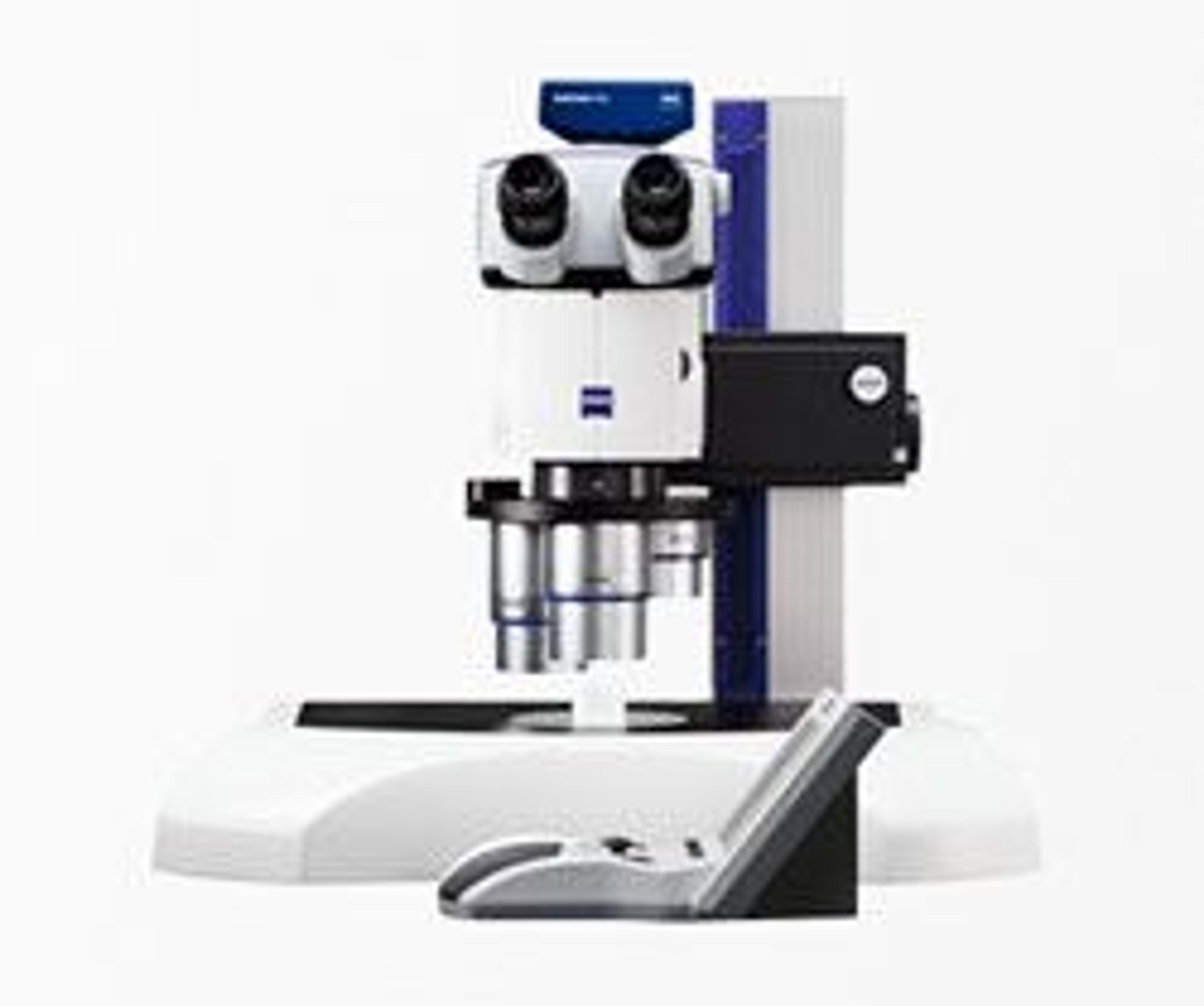
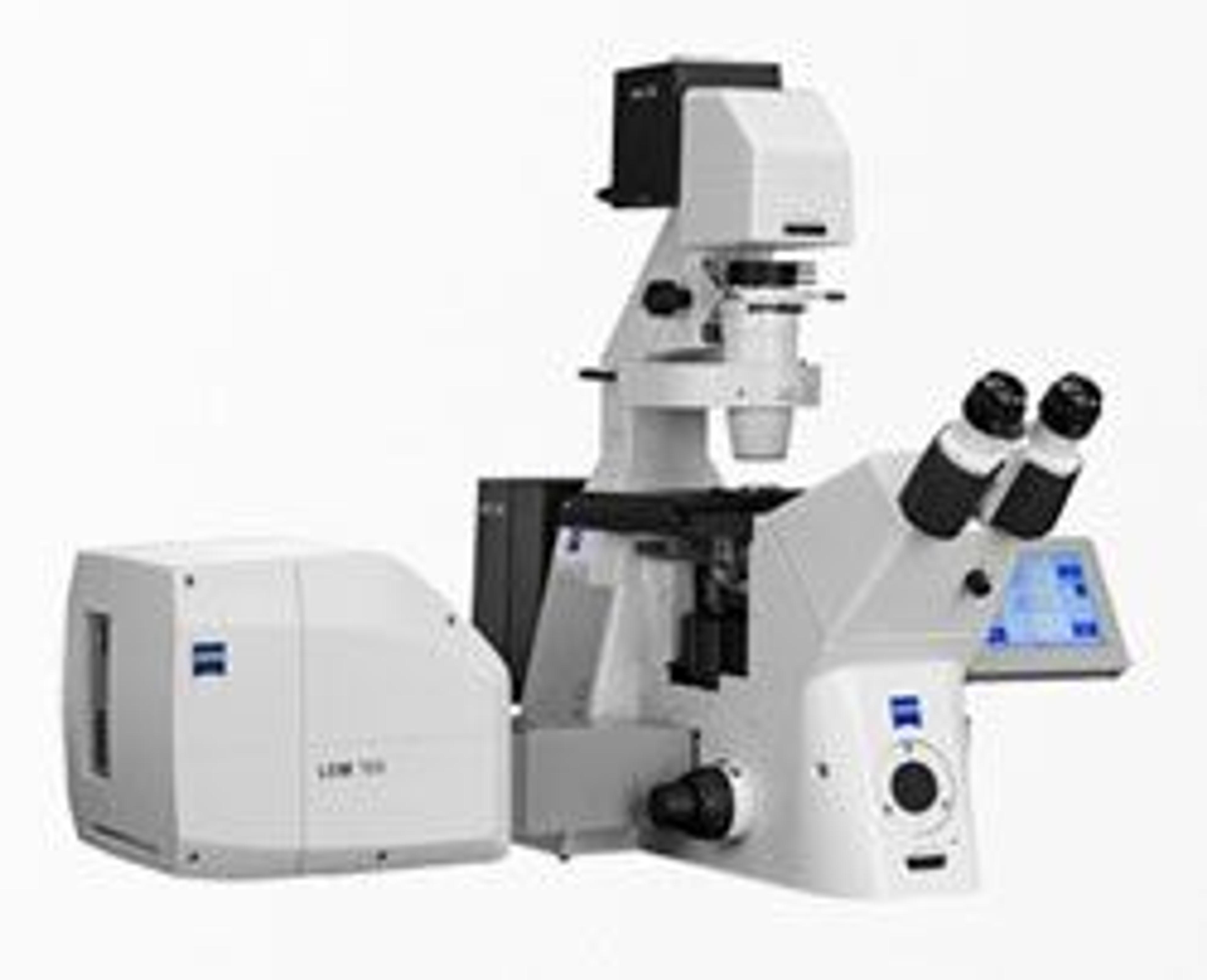
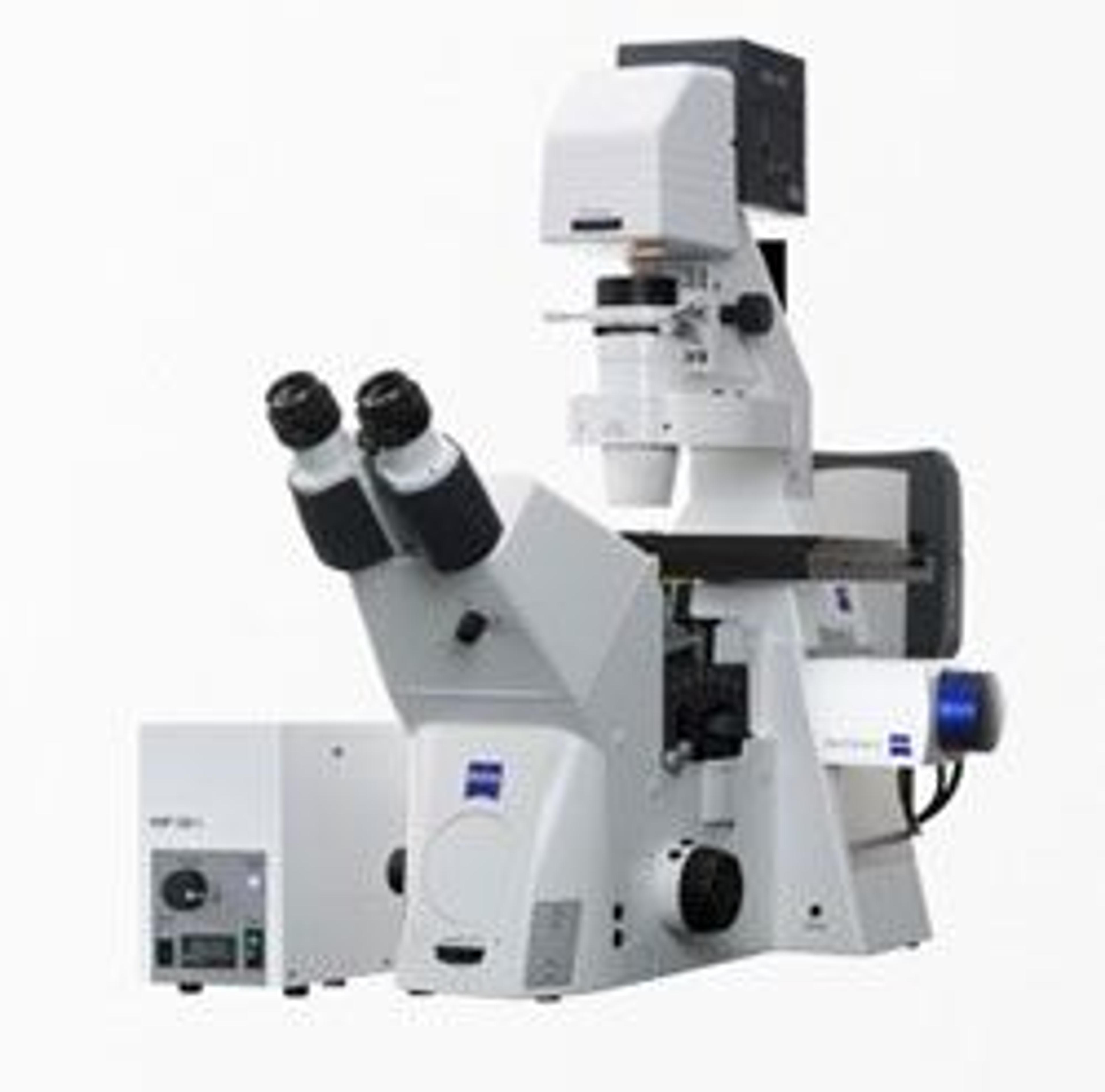
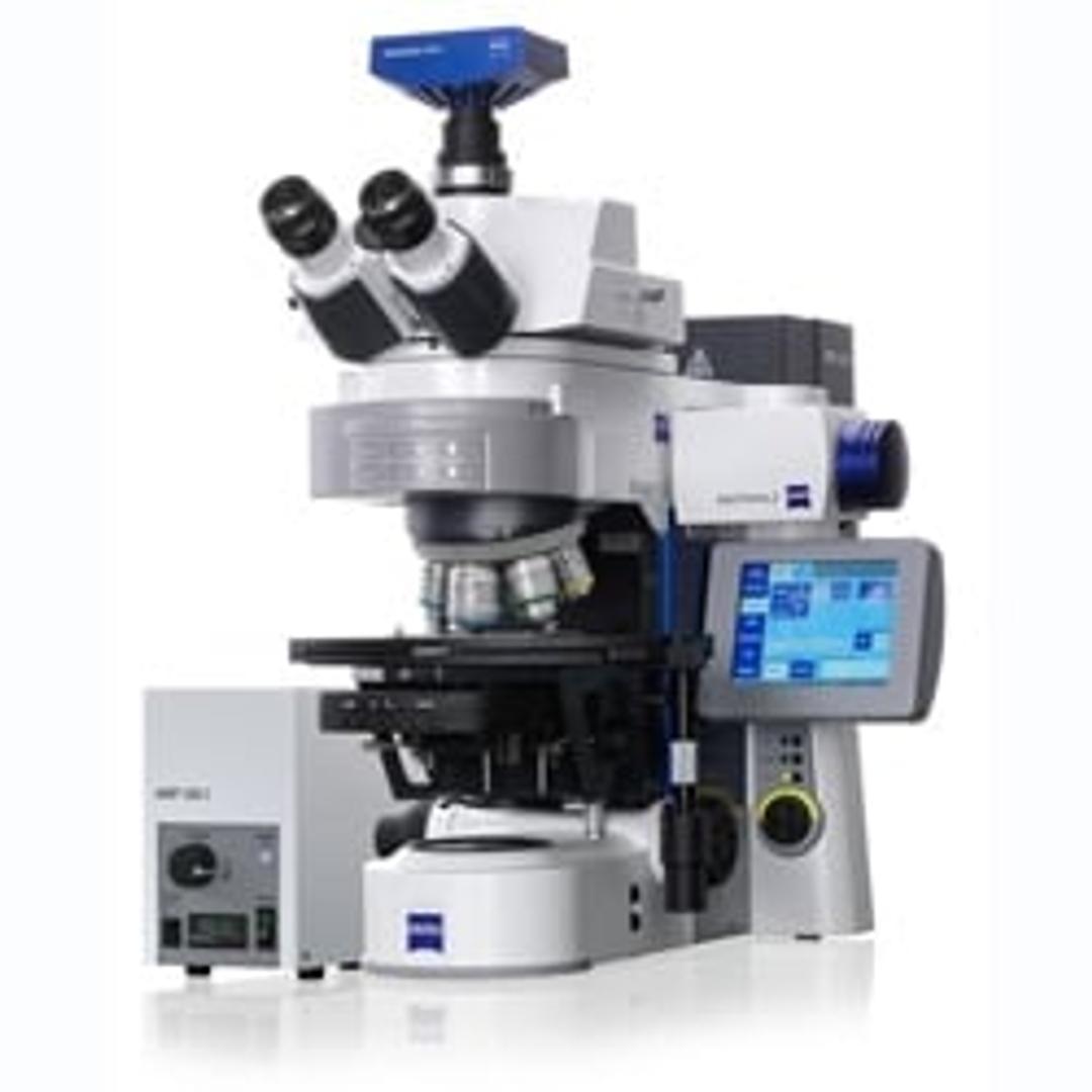
ZEISS ApoTome.2
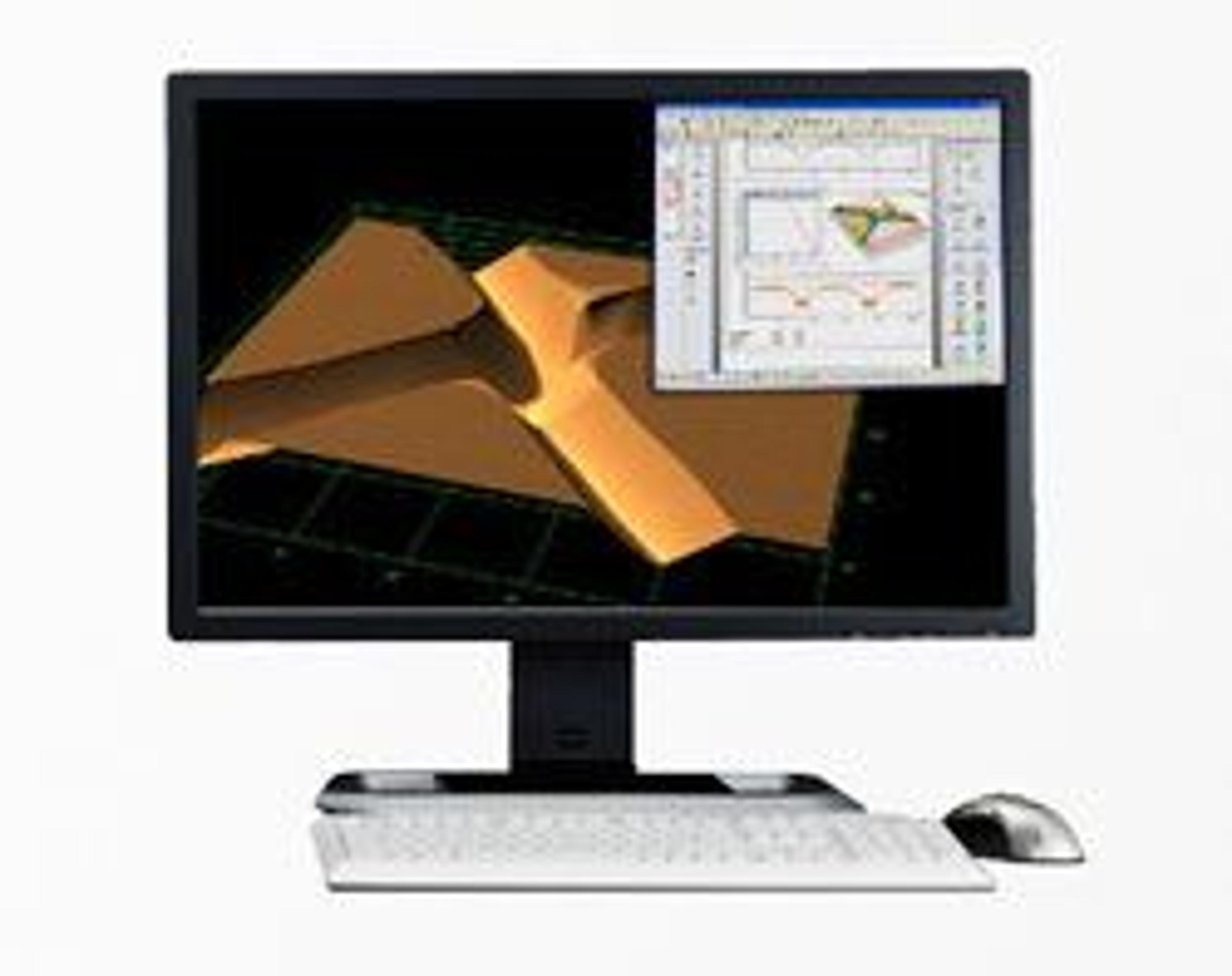
Optical Sections in Fluorescence Imaging.


Receive your quote directly from the manufacturer.
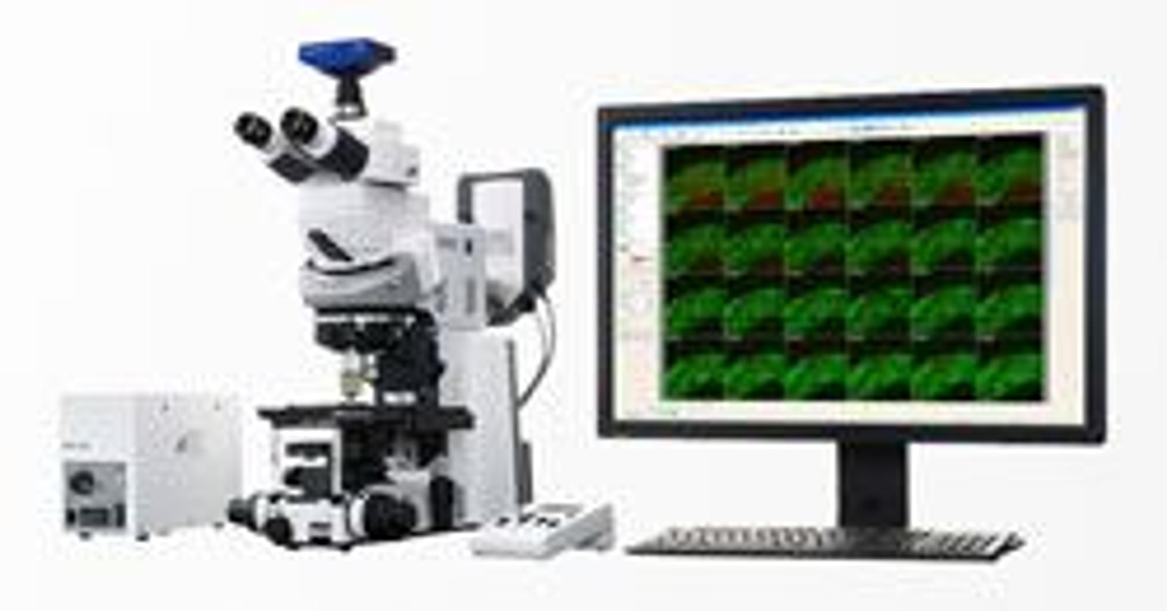
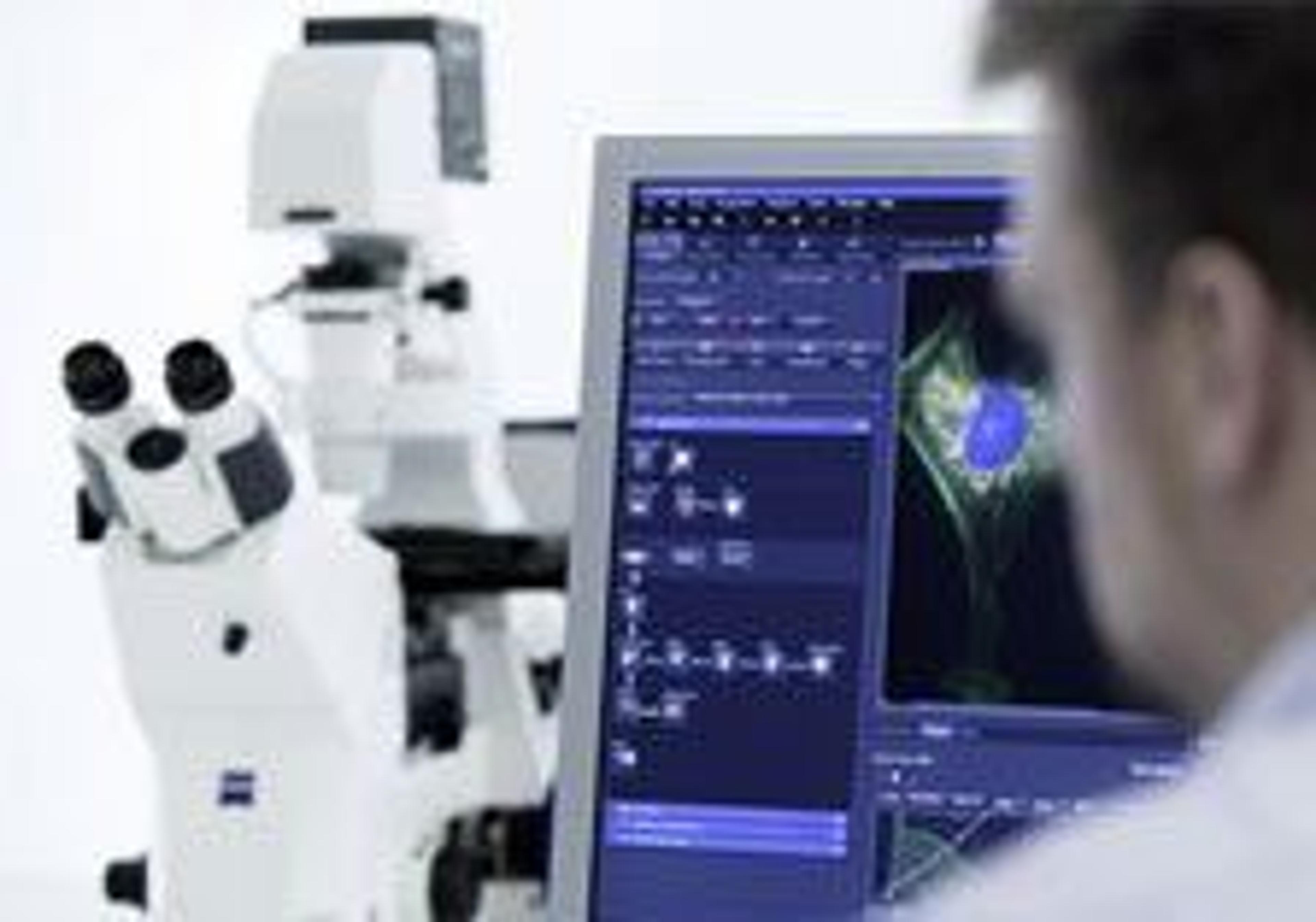
Fluorescent imaging
The Apotome2 makes imaging easy. The images are crisp and clean. Converting the images to tiff or jpeg is really easy. I like that I can make final adjustments to the contrast and brightness to get the best image possible.
Review Date: 31 Mar 2015 | ZEISS Research Microscopy Solutions
Imaging
I wouldn't say easy to use but it does gather publication quality images.
Review Date: 26 Mar 2015 | ZEISS Research Microscopy Solutions
Indispensable for when you want a precision photomicrography with different levels in a single plane!
Review Date: 28 Nov 2014 | ZEISS Research Microscopy Solutions
Imaging
Apotome is fairly easy to use and easy to set up. It provides excellent resolution in making stacks of thin slices stained with fluorescent proteins.
Review Date: 19 Nov 2013 | ZEISS Research Microscopy Solutions
Apotome is great for doing stacks but a confocal is much easier to use.
Review Date: 5 Jan 2011 | ZEISS Research Microscopy Solutions
Good product.
Review Date: 11 Jan 2008 | ZEISS Research Microscopy Solutions
Review Date: 10 Jan 2008 | ZEISS Research Microscopy Solutions
Create optical sections of your fluorescent samples – free of scattered light. With structured illumination, you know that only the focal plane appears in your image: ApoTome.2 recognizes the magnification and moves the appropriate grid into the beampath.
The system then calculates your optical section from three images with different grid positions without time lag. It’s a totally reliable way to prevent scattered out-of-focus light, even in your thicker specimens.
Yet your system remains just as easy to operate as always. You get images with high contrast in the best possible resolution – simply brilliant optical sections.