NutriFreez™ D10 Cryopreservation Medium
Biological IndustriesA chemically defined, animal component-free, protein-free cryopreservation solution
Products, services, reviews and techniques used in the identification and validation of the disease-causing target genes and proteins.
A chemically defined, animal component-free, protein-free cryopreservation solution
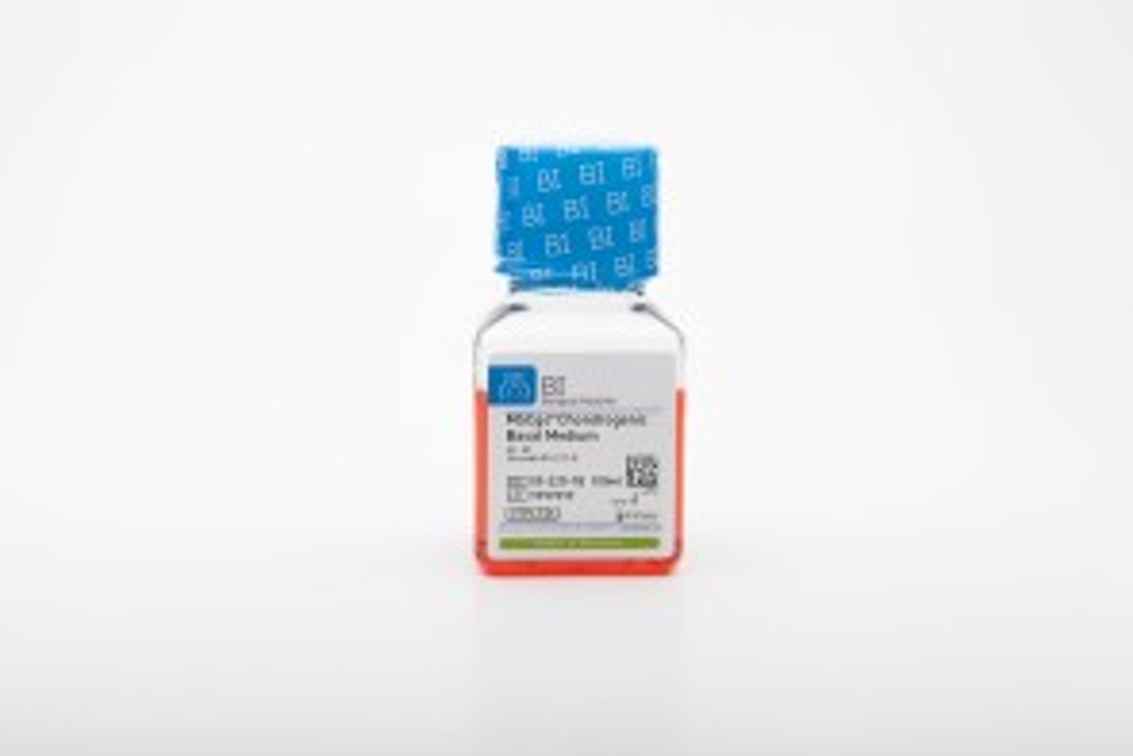
Complete, serum-free, xeno-free, ready-to-use kit, Optimized for directed differentiation of hMSCs to chondrocytes. To be used with MSCgo Chondrogenic Differentiation Supplement Mix (05-221-1D)
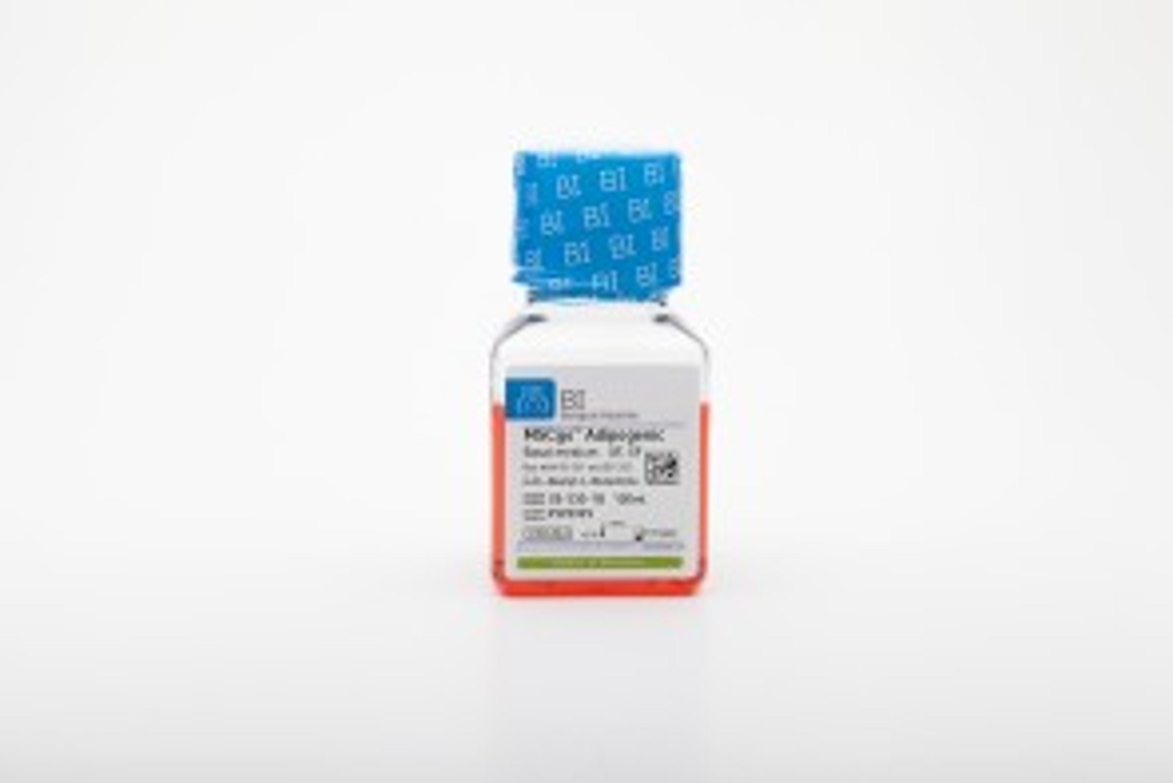
Complete, serum-free, xeno-free, ready-to-use kit. To be used with MSCgo™ Adipogenic Supplement Mix I (05-331-1-01) and MSCgo™ Adipogenic Supplement Mix II (05-332-1-15) to make the complete kit optimized for directed differentiation of hMSCs to adipocytes.
Defined, xeno-free, serum-free medium for long-term expansion of large and small vessels of endothelial cells. To be used with EndoGo™ XF Supplement Mix ( 05-410-1-25). *Requires addition of 2 to 5% of OTC human AB serum or platelet lysate.
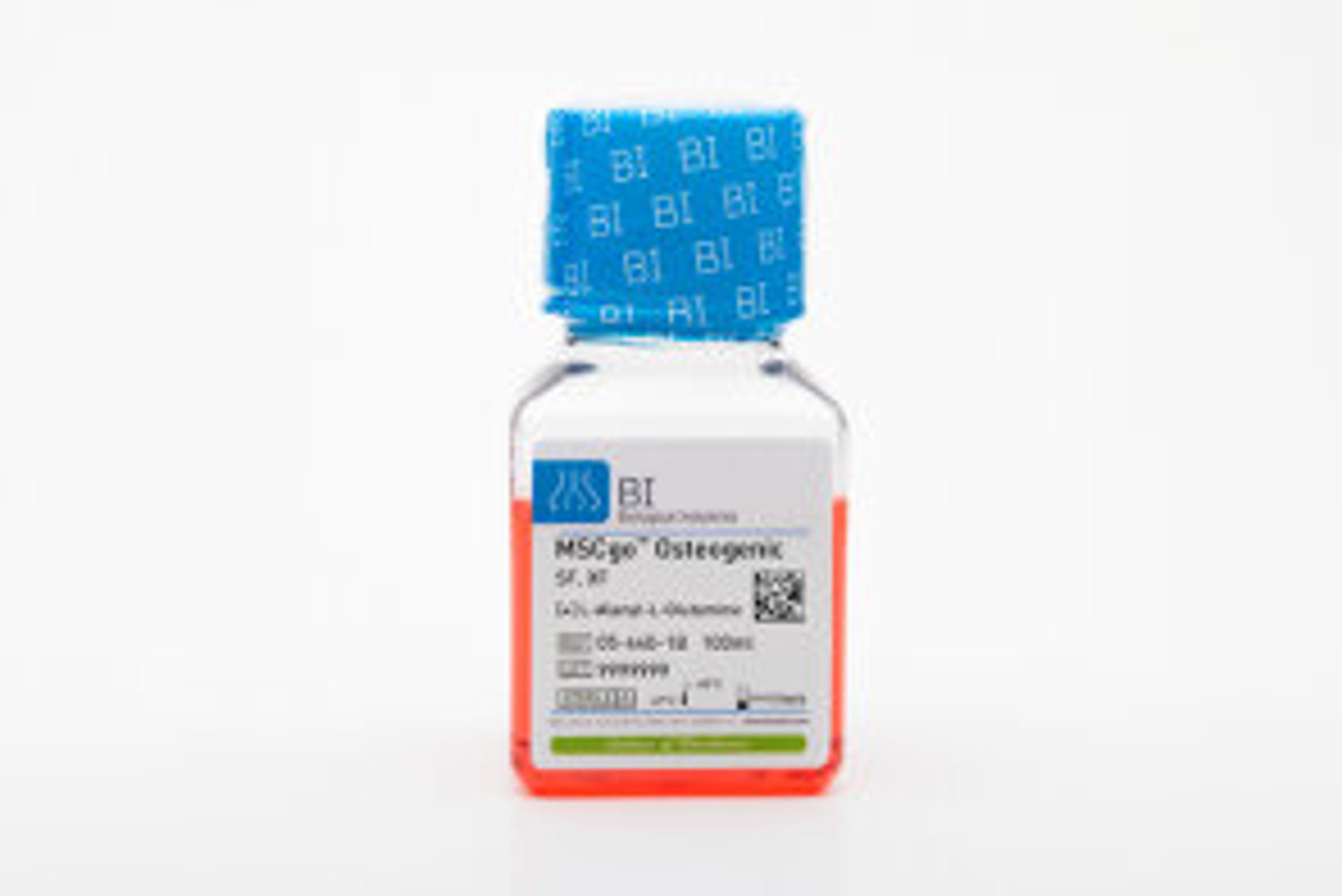
Complete, serum-free, xeno-free, ready-to-use medium, Optimized for directed differentiation of hMSCs to osteoblasts
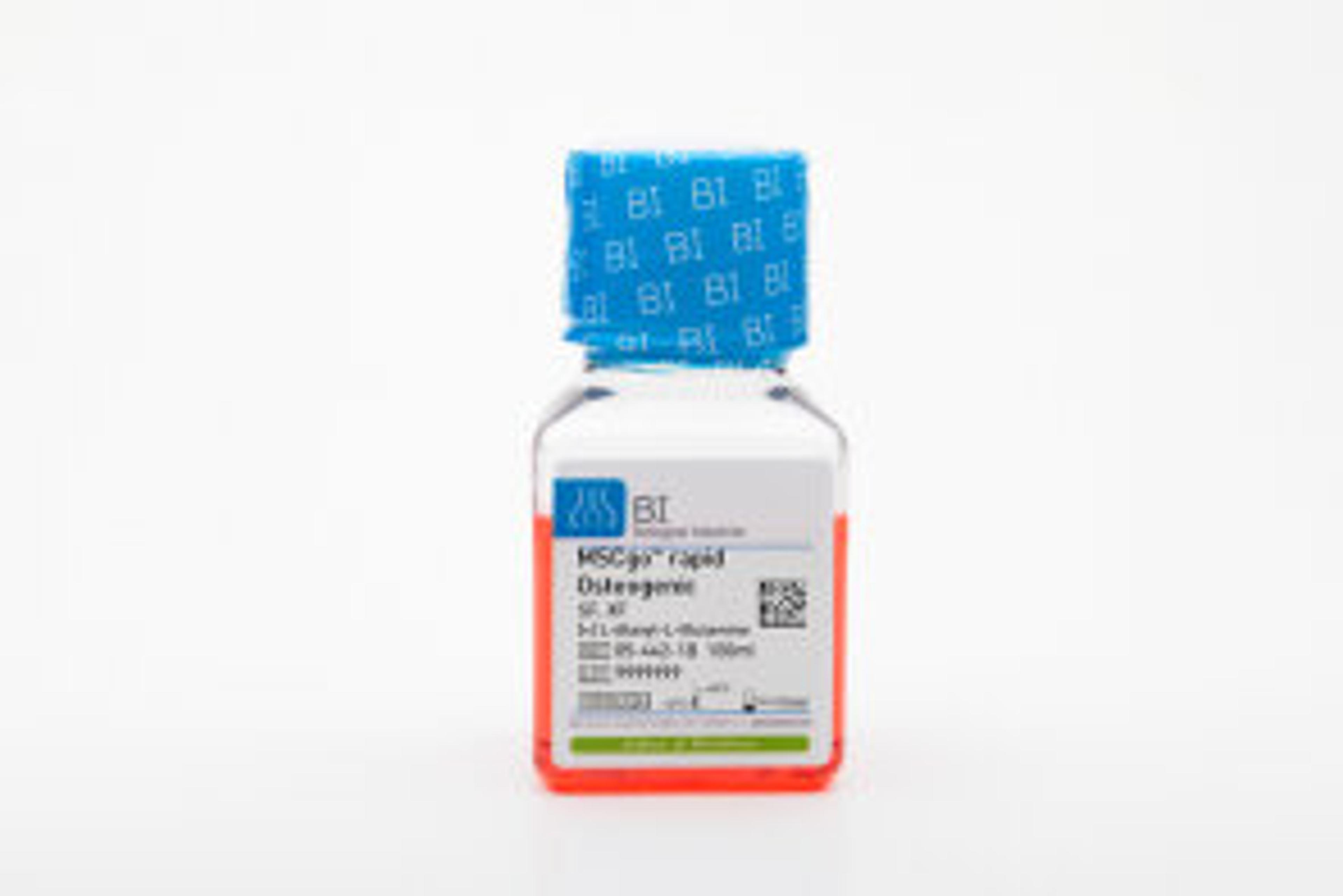
Complete, serum-free, xeno-free, ready-to-use medium, Optimized for faster directed differentiation of hMSCs to osteoblasts
Fibronectin is an attachment factor that facilitates the attachment and cytoplasmic spreading of all types of anchorage-dependent cells.
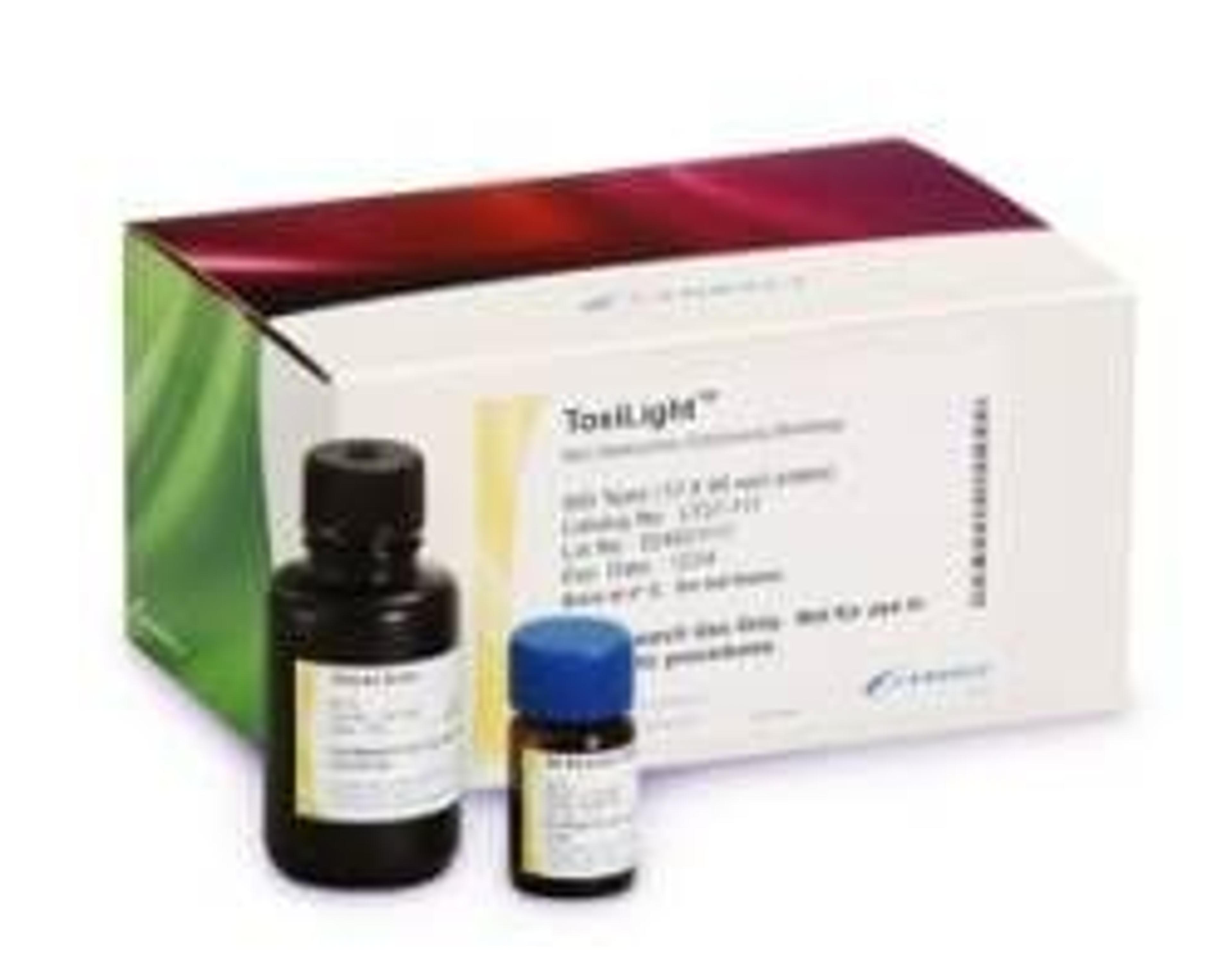
The ToxiLight® BioAssay Kit is a bioluminescent, non-destructive cytolysis assay kit designed to measure the release of the enzyme, adenylate kinase (AK), from damaged cells. The ToxiLight BioAssay Kit also facilitates high content screening by allowing other tests to be performed on the original cells For example, the ToxiLight BioAssay Kit is fully compatible with the ReportaLight™ BioAssay Kit, thus facilitating measurement…
Xeno-free, for attachment and spreading of hMSCs cultured in serum-free conditions
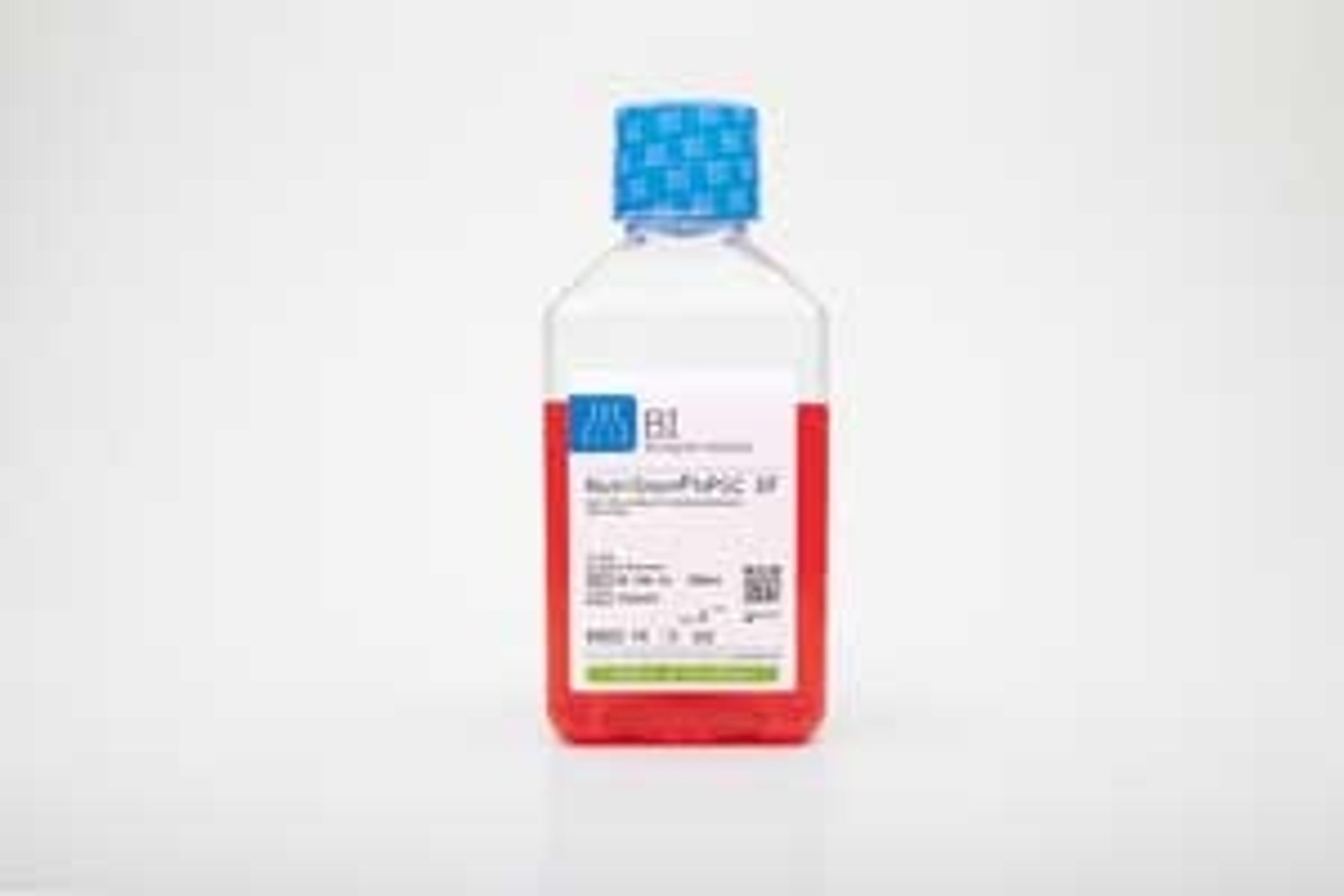
Defined, xeno-free, serum-free medium, Designed for optimal growth and expansion of human iPS and hES cells.