ResourceLife Sciences
14-plex sample imaging of liver cancer modalities using one-step staining and imaging
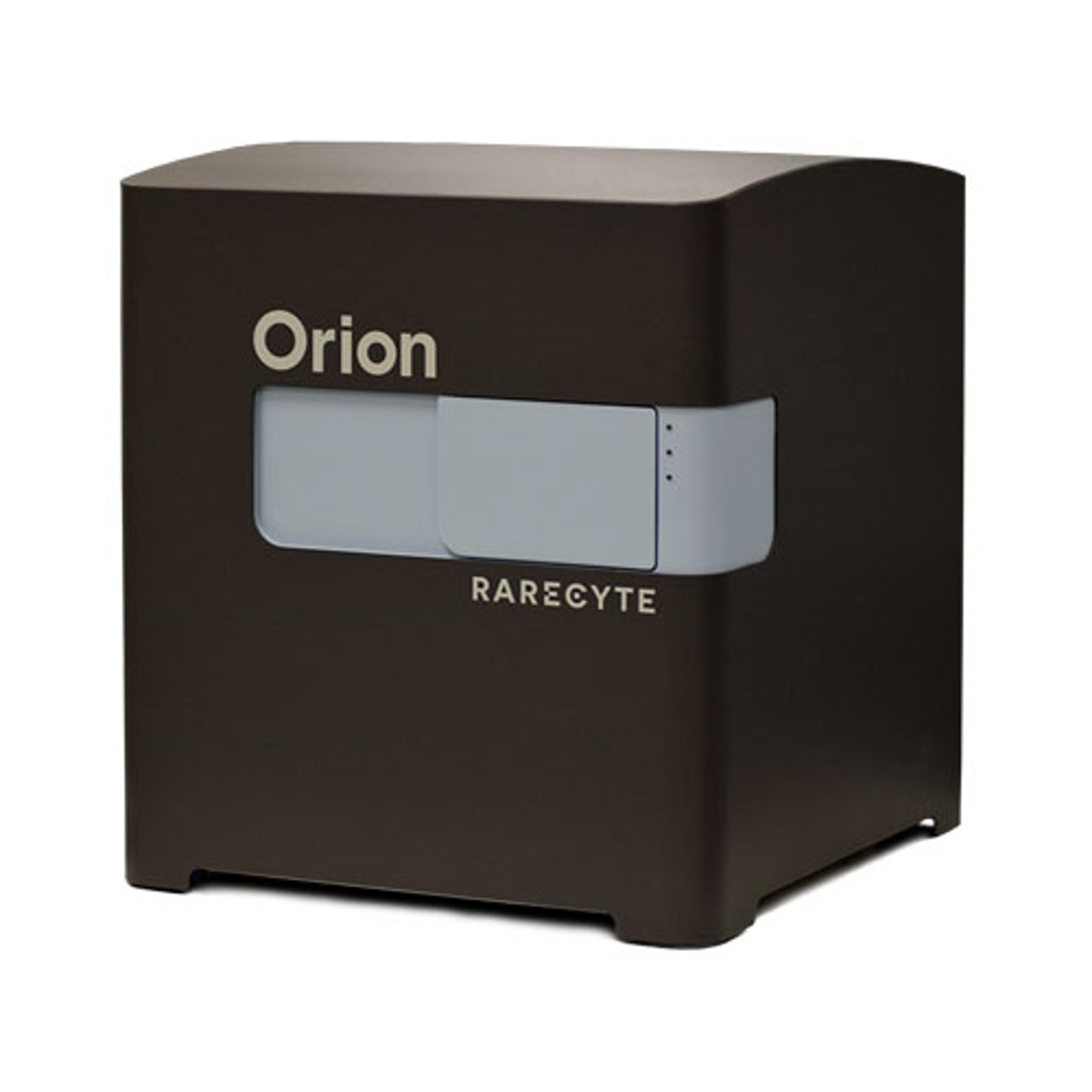
17 Jun 2024RareCyte presents a study of a tumor microenvironment, using the Orion™ imaging system. The formalin-fixed paraffin-embedded (FFPE) liver section was stained using a 14-plex immunofluorescence (IF) panel in one staining round, followed by whole-slide imaging with the Orion instrument, allowing for the traditional pathology analysis to be complimented by same-cell phenotyping. The liver section exhibits extensive infiltration by metastatic moderately-differentiated adenocarcinoma with high Ki-67 proliferation index and central dirty necrosis morphologically consistent with primary tumor from the colon.

