ResourceLife Sciences
Automated hemocytometer-based live/dead Cell Counting using hase contrast and color brightfield imaging
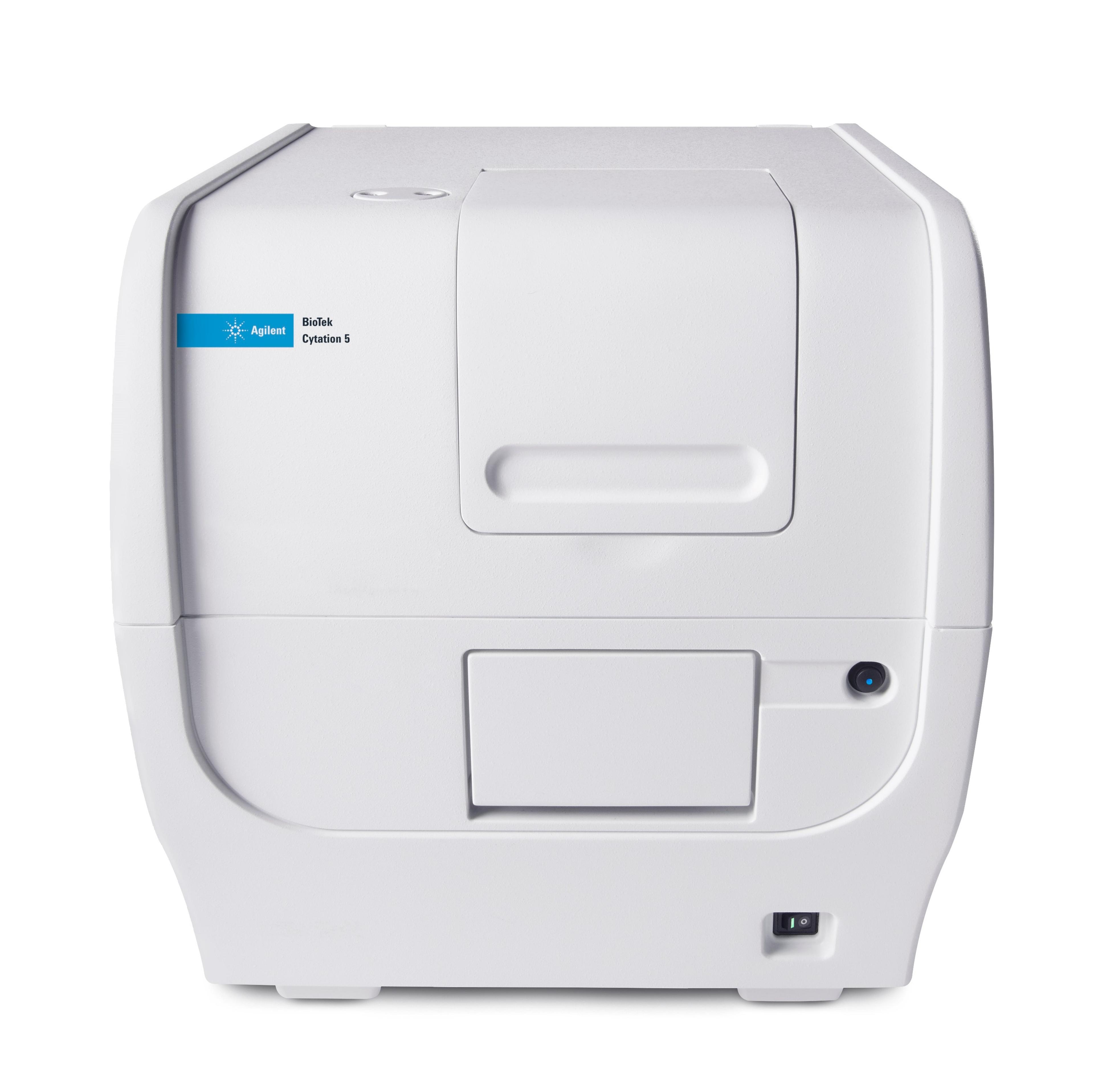
13 Jul 2020In this application note, BioTek demonstrates how the hemocytometer cell counting process can be automated using a Cell Imaging Multi-Mode Reader equipped with contrast enhancement technologies. For total cell counts, the Cell Imager uses automated phase-contrast microscopy to count the total number of cells in the field of view which is about 2.9 times larger than the total ruled grid area. This serves to improve the counting statistics relative to counting manually. Furthermore, cell viability can also be determined through the use of automated color imaging of tryptan blue-stained cells in the same field of view.

