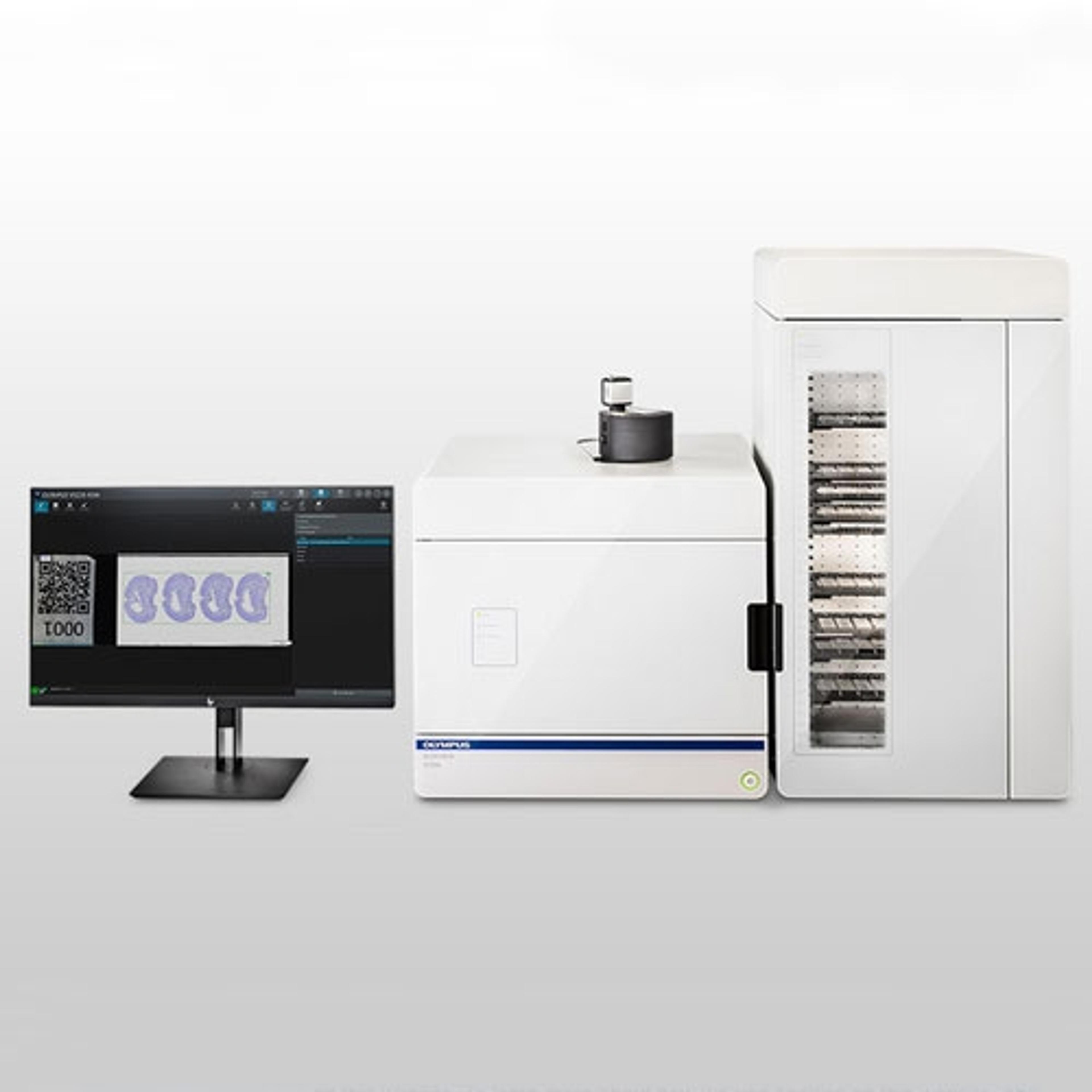
Digitizing very thick fluorescent samples with the SLIDEVIEW VS200 research slide scanner
7 Dec 2020In a recently published application note, Olympus demonstrates the uses and benefits of the SLIDEVIEW VS200 research slide scanner. The document details a case study from the Max Planck Institute of Molecular Cell Biology and Genetics (MPI-CBG) in Dresden, Germany, where researchers are investigating the regeneration capabilities of the planarian flatworm Schmidtea mediterranea.
With low associated costs and a recently sequenced genome, S. mediterranea is a popular model for studying the biology of system regeneration. However, the specimens can be relatively thick, measuring approximately 200-300µm. Researchers from MPI-CBG are using the VS200 slide scanner to acquire high-resolution fluorescence images of these specimens.
The Olympus VS200 comes equipped with TruSight Live and Z-stack functionality, which reduce diffused light from the areas above and below the focus plane. A special 2D deconvolution algorithm then recalculates image data, creating a sharper and clearer result. After image acquisition, users can seamlessly focus through the Z-stack to see the signal distribution through the sample thickness. These options are essential for visualizing thicker specimens that may be subject to blurring.
The application note also contains comparative images to demonstrate the benefits of using the Olympus VS200. The images depict multiple differentiated fluorescent markers (DAPI, FITC, and CY3), which allows for visualization of various structural features of S. mediterranea in fine detail.