ResourceLife Sciences
Fluorescent Image Analysis – Cell Division in Spheroids
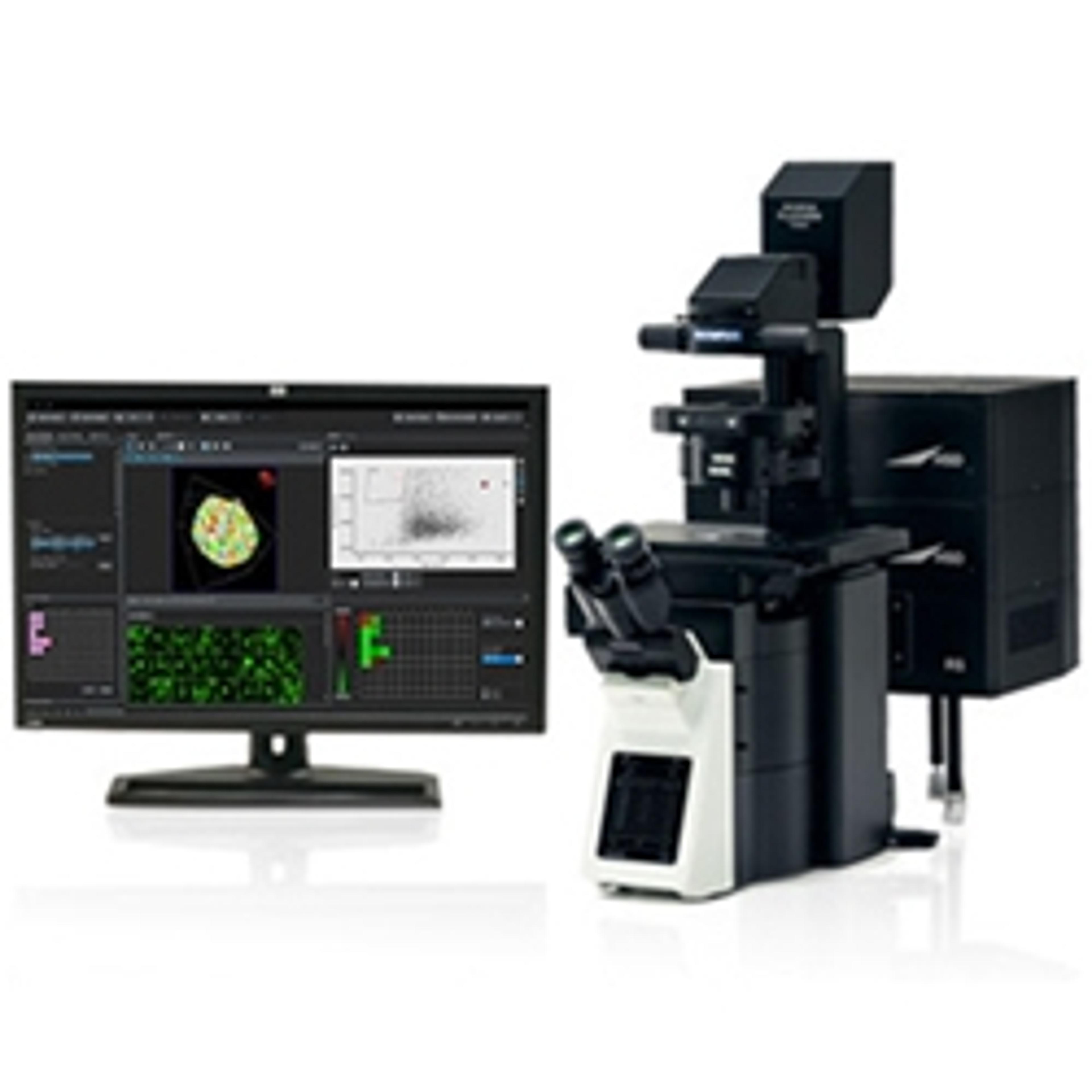
6 Jun 2019Cell division inside spheroids can be visualized by staining cell nuclei and microtubules. The number of cells in mitosis in spheroids can be quantitatively evaluated using images captured by a laser confocal microscope and NoviSight™ software's counting module.