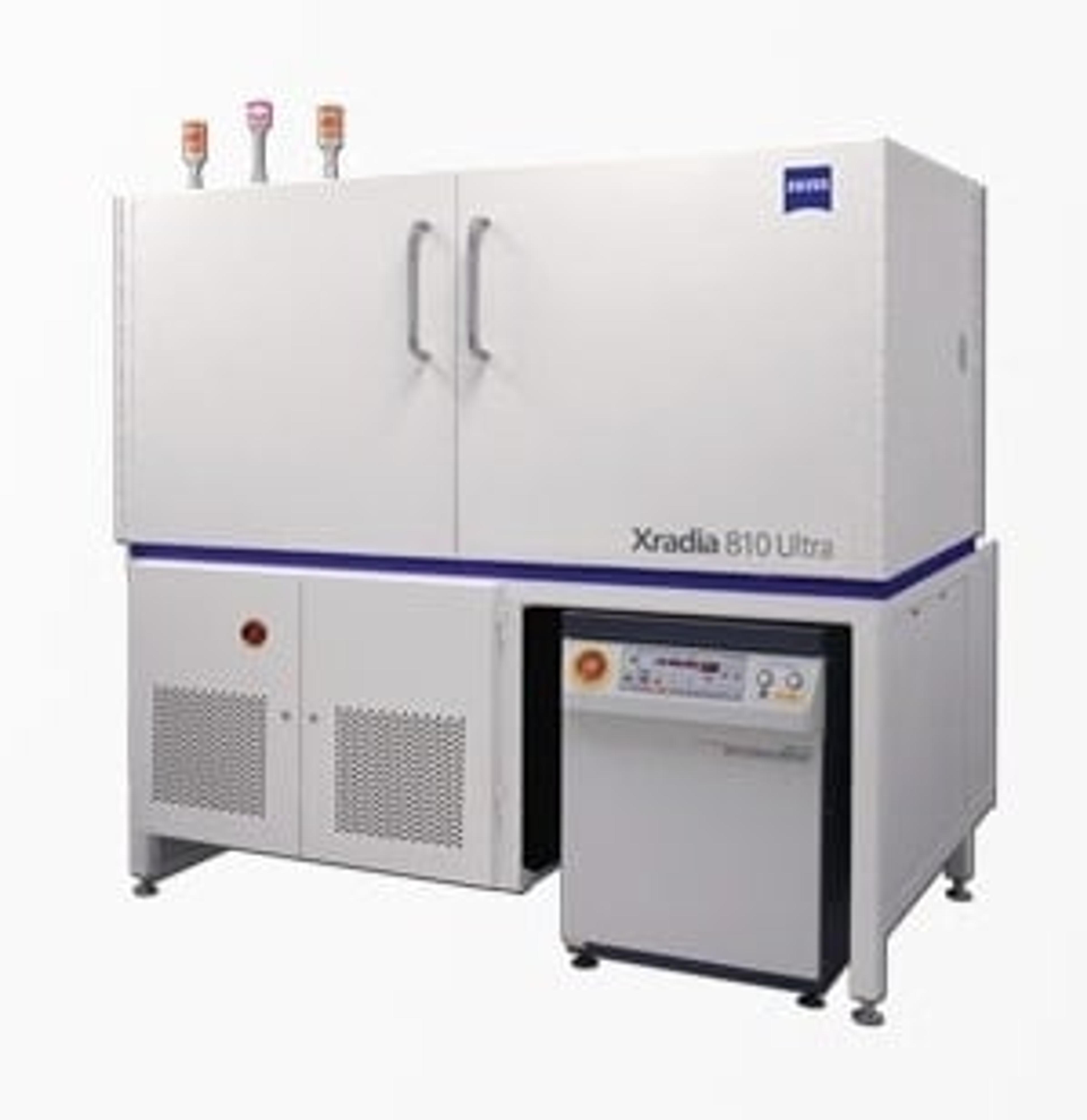
In situ 3D imaging of crack growth in dentin at the nanoscale
14 Jun 2024Dentin is a nano-composite material which forms the majority of the mineralised tissue in teeth. A better understanding of fracture in dentin is important to develop a framework for failure prediction, not only for clinical understanding but also for developing biomimetic restorative materials that are able to mimic the tissue’s mechanical response. ZEISS shows how a novel in situ nanomechanical test stage for the ZEISS Xradia Ultra nanoscale X-ray microscope could be used to initiate and propagate cracks in elephant dentin (tusk) during tomography. This enabled the progressive crack growth to be studied in 3D and in situ (under load) at 150 nm resolution for the first time. The results can provide new insights into anisotropic fracture behavior and crack shielding mechanisms.

