ResourceLife Sciences
Quantitative analysis of colorectal adenocarcinoma images
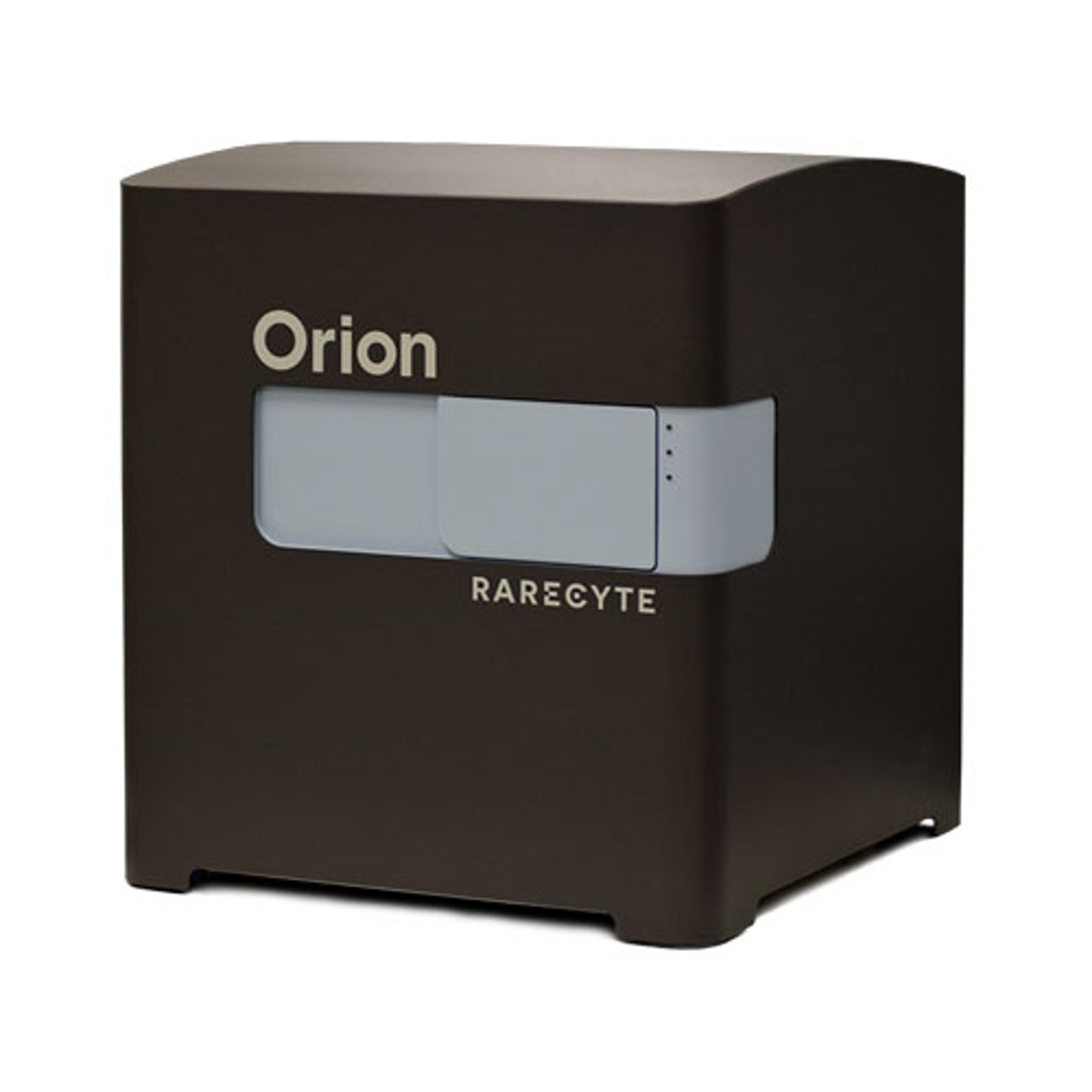
17 Jun 2024Understanding the tumor microenvironment is particularly important for oncology studies. RareCyte demonstrates how the Orion™ spatial biology platform has been used to investigate a sample of invasive colorectal adenocarcinoma using whole slide, single-step high-plex staining and imaging at single-cell resolution followed by quantitative analysis. The data reveal a distinction between normal colonic epithelium, well-differentiated adenocarcinoma with immune cell collection, and an infiltrating border of the carcinoma. These data also highlight the importance of sufficient plex, resolution and whole slide context to derive reliable spatial biomarkers of potential prognostic value.

