Product NewsLife Sciences
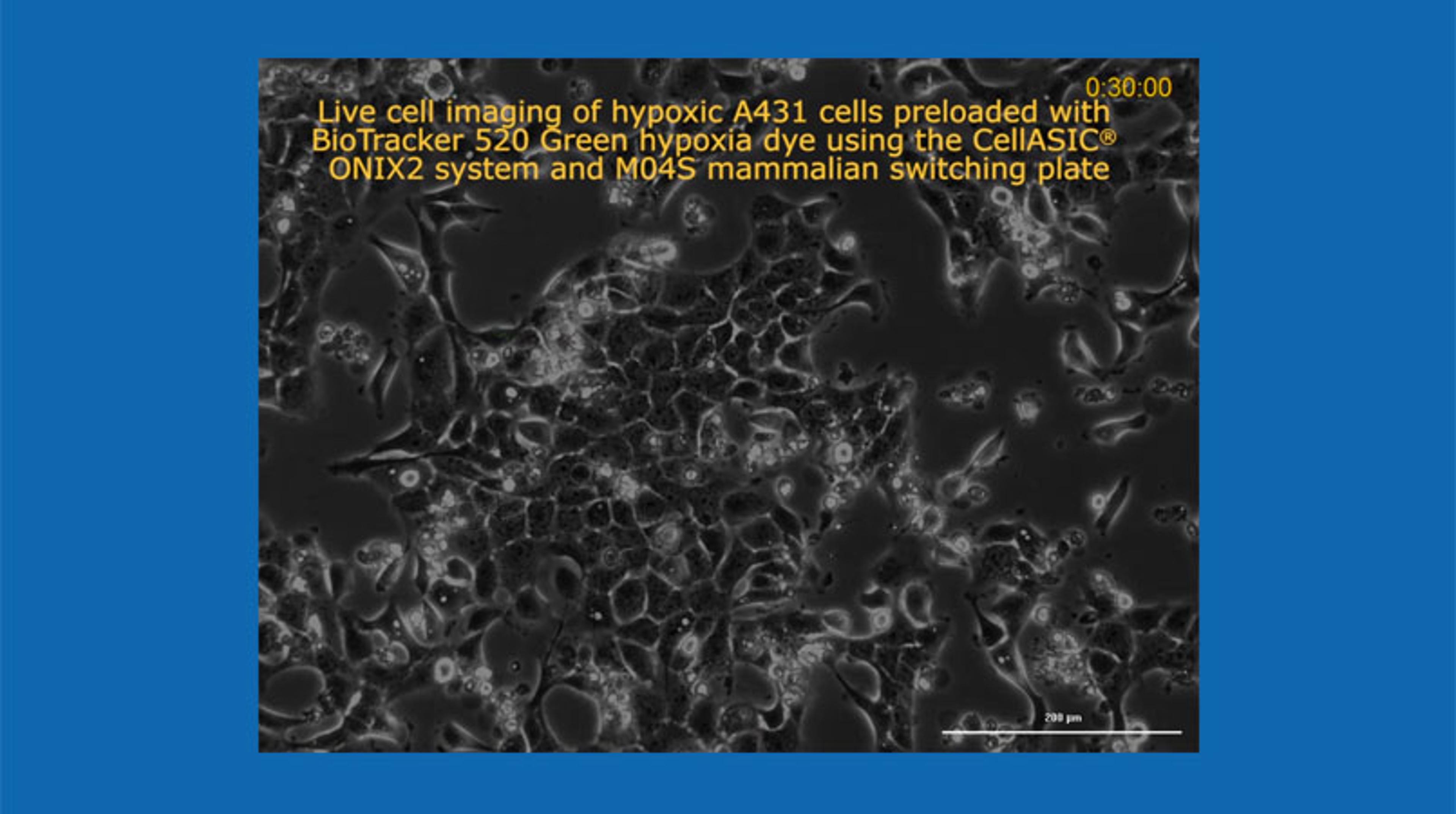
Live Cell Imaging of Hypoxia in Cancer Cells using the CellASIC® ONIX2 Microfluidic System
14 Oct 2019
A431 epidermal carcinoma cells were incubated 16 hours overnight with 3µM BioTracker 520 Green Hypoxia Dye in complete DMEM by gravity perfusion at 37°C under normoxic conditions (20% O2) using the CellASIC® ONIX2 System. After overnight incubation, cells were perfused with complete DMEM media without dye at 1 psi under hypoxic conditions (0% O2) for 16 hours at 37°C. Fluorescent images were captured over 16 hours using 465 nm excitation, and a 525 nm emission filter. The BioTracker 520 Green Hypoxia Dye increases in fluorescence intensity with decreasing oxygen level.
