WebinarLife Sciences
Applications of high-content imaging in 3D models of disease
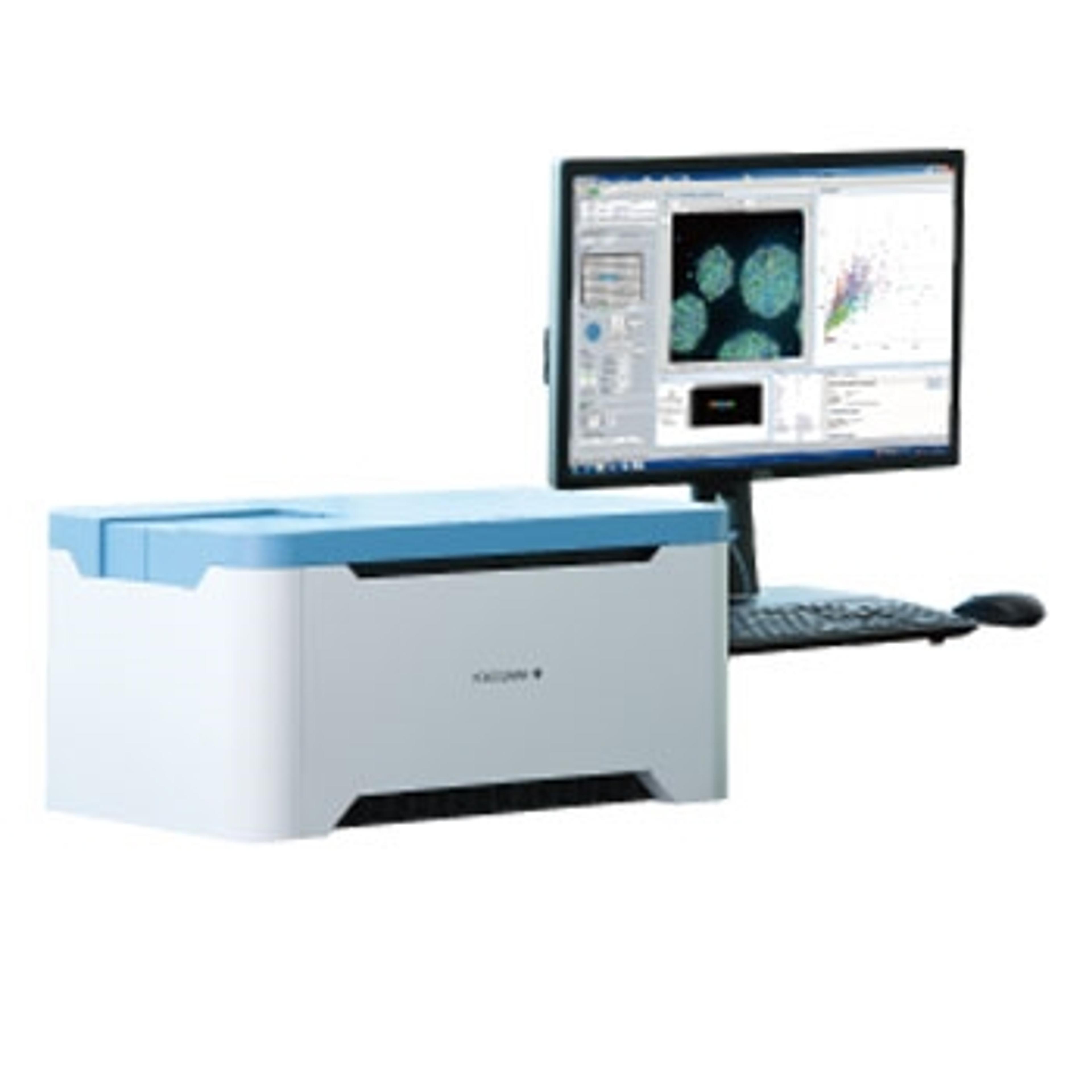
Generating translatable high-content imaging data from physiologically-relevant cell models, including 2D and 3D structures, is extremely valuable for drug discovery and pre-clinical research. In this webinar, James Evans, CEO of PhenoVista Biosciences presents case studies on how Yokogawa’s Benchtop CQ1 Confocal System can improve throughput and standardize processes for complex 3D cell-based phenotypic assays.
Key learning objectives:
- Strategies for designing and implementing high-content screening assays
- Approaches for deciding between 2D and 3D model systems
Who should attend?
Scientists and researchers from industry and academic backgrounds who are interested in high-content imaging and specific applications including:
- Living-cell imaging
- Organoids and spheroids
- 3D high-content imaging
- Organ on a chip
- Microfluidics
- Cell painting
- Drug screening/discovery
Speakers

CEO, PhenoVista Biosciences

Associate Editor, SelectScience

