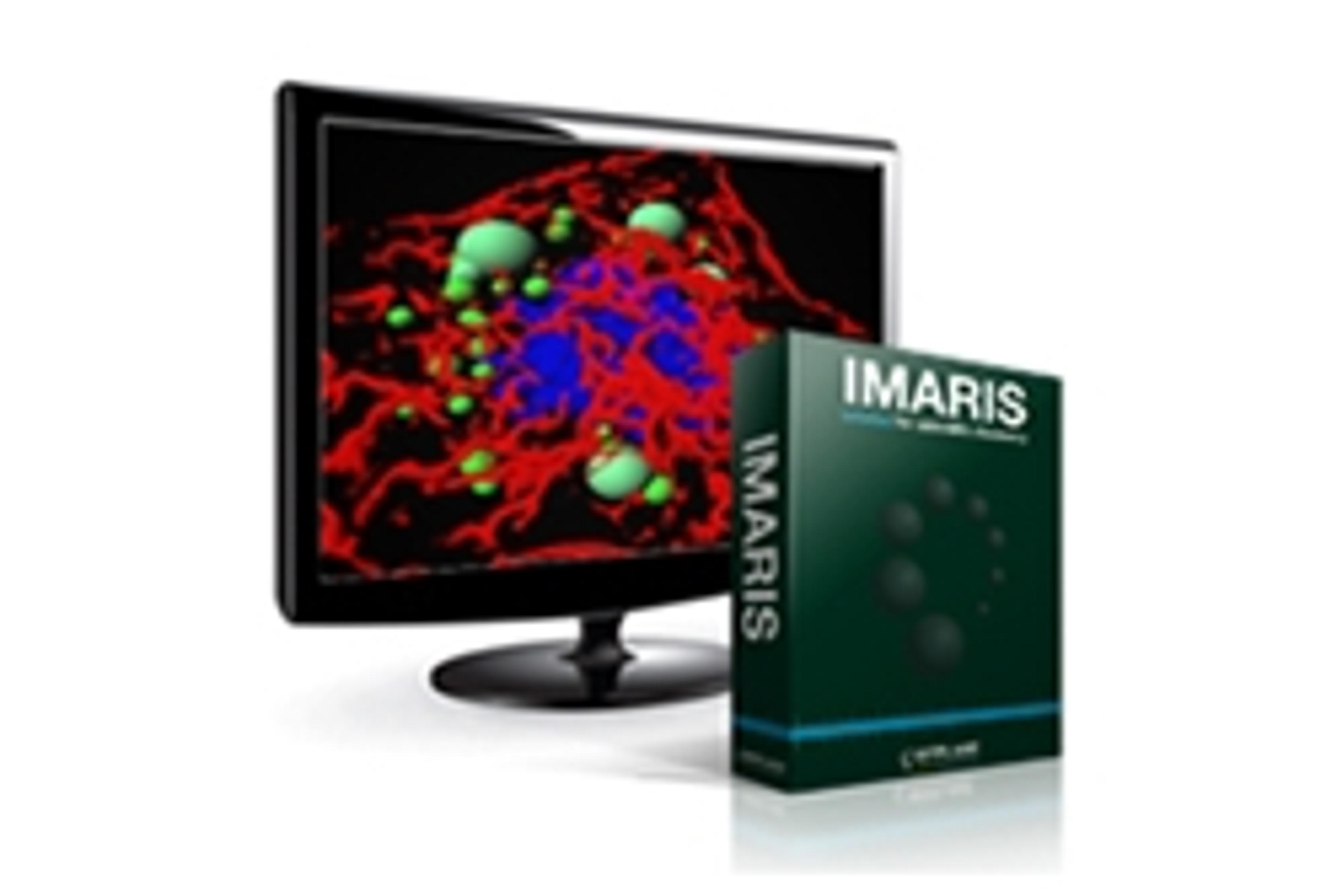
Combining fluorescence microscopy and 3D reconstruction to investigate neuron-glia interaction in different neurological diseases
Dr. Danielle Beckman, Project Scientist at the California National Primate Research Center, will introduce how they apply high-resolution confocal microscopy to help the development of different primate models for neurological disorders. She will also show how acquisition of z-stack images in association with 3D reconstruction performed at Imaris helps them investigate neurodegeneration and neuroinflammation in different disease models.
Key learning objectives
- Microglia morphology: Learn how to create and edit 3D surfaces to analyse microglia volume and interaction with other cell types. Understand what the indicators are for microglia activation and neuroinflammation.
- Viral infections: Discover how to identify and quantify different markers for viral infection.
- Neuronal death: Understand how to detect neurons dying using multilabel confocal microscopy, especially in combination with neuroinflammatory markers
Who should attend?
Both new and experienced Imaris users interested in accelerating microscopy projects. The research is relevant across life science projects where visualization and analysis is required.
Certificate of attendance
All webinar participants can request a certificate of attendance, including a learning outcomes summary, for continuing education purposes.
Speakers
Danielle Beckman has a Ph.D in neuroscience and currently works as Project Scientist at the California National Primate Research Center. She is particularly interested is applying high-resolution microscopy to investigate early biochemical events in the brain that lead to neuroinflammation and neurodegenerative processes.
Moderator