Size and fluorescence calibrated imaging flow cytometry: From arbitrary to standardized units
Thursday, November 20, at 15:00 GMT | 16:00 CET | 10:00 EST | 7:00 PST
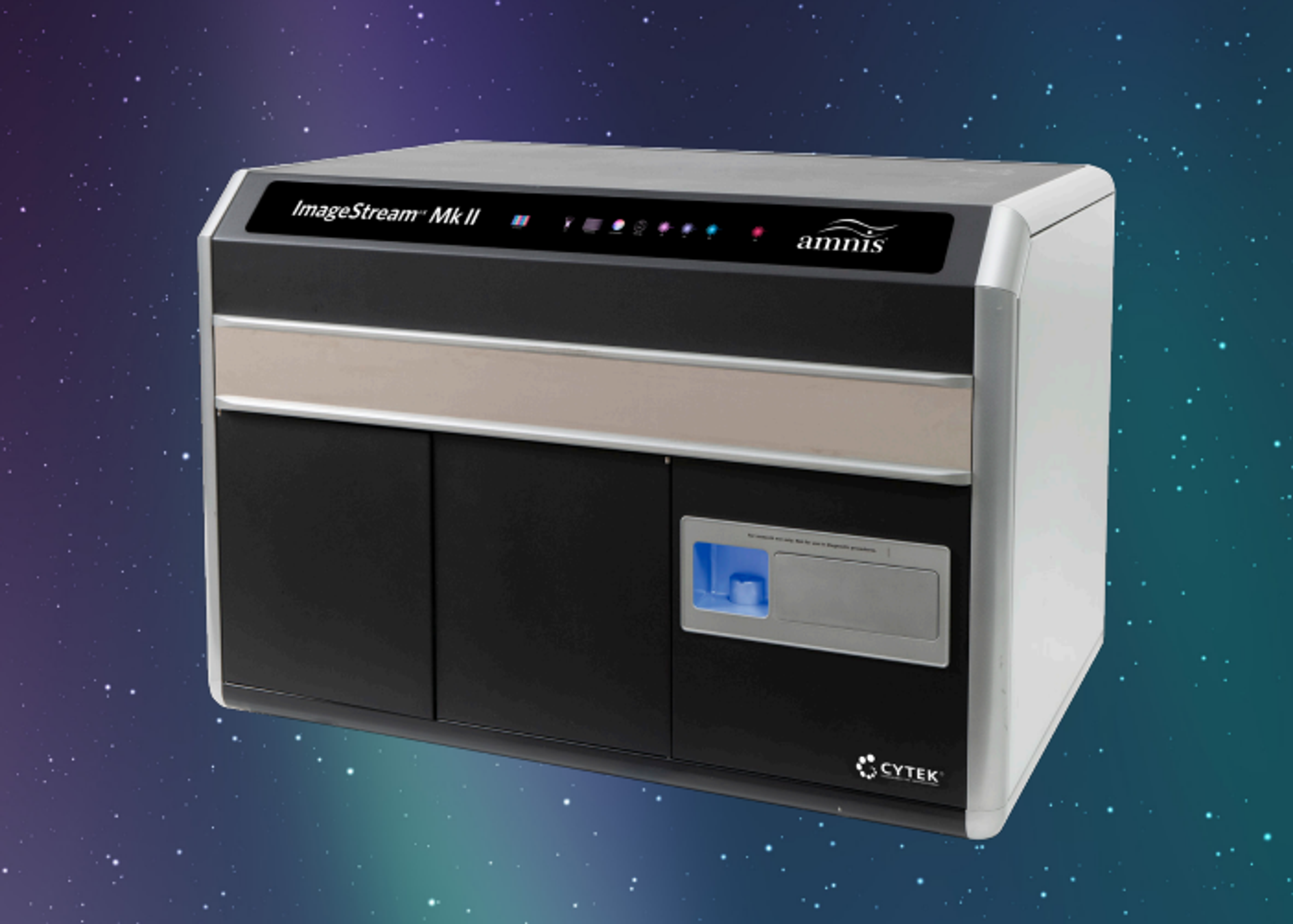
Imaging flow cytometry (IFC) has emerged as a powerful approach for single extracellular vesicle (EV) characterization, yet its broader adoption has been hindered by challenges in assay standardization and data comparability. This webinar will highlight recent methodological advances addressing these limitations. Building on earlier work that established workflows for direct detection and phenotyping of EVs in unprocessed plasma, current efforts focus on developing rigorous calibration strategies for IFC.
Recent work has introduced bead-based size calibration and fluorescence intensity standardization, enabling instrument-specific signals to be expressed in quantitative units. These approaches markedly improve reproducibility across different platforms and laboratories, thereby supporting cross-study comparability. Standardized IFC data further provide a framework for quantitative analyses in both fundamental EV research and the quality assessment of vesicle-based therapeutics.
Together, these methodological advances position IFC as a calibrated, quantitative platform for nanoparticle cytometry, with the potential to accelerate both biomarker discovery and translational applications in extracellular vesicle science.
Topics discussed in this webinar will include:
- Advances in EV assay standardization and data comparability
- Development of calibration strategies for IFC
- Introduction of bead-based standardization methodology for quantitative EV analyses
Who should attend?
- Researchers using IFC who are currently or wish to perform small particle analysis
- Anyone interested in flow cytometry applications and small particle analysis
Certificate of attendance
If you attend the live webinar, you will automatically receive a certificate of attendance, including a learning outcomes summary, for continuing education purposes.
If you view the on-demand webinar, you can request a certificate of attendance by emailing editor@selectscience.net.
For Research Use Only. Not intended for use in diagnostic procedures.
Speakers
Dr. Wouter W. Woud is an expert in nanoparticle flow cytometry, with a particular focus on imaging flow cytometry for the study of extracellular vesicles. Wouter obtained his Ph.D. at the Rotterdam Transplant Institute, Erasmus MC, The Netherlands, where he developed an imaging flow cytometry-based method to detect and characterize single vesicles directly in human plasma. His doctoral thesis, “Fantastic vesicles and how to find them” (2023), outlined best practices for instrument calibration, assay design, and data analysis, helping to establish imaging flow cytometry as a reliable tool for vesicle research. During his Ph.D. studies, Dr. Woud demonstrated how imaging flow cytometry can uncover clinically relevant vesicle populations without the need for complex pre-isolation steps. More recently, he helped introduce calibration methods that make results comparable across different instruments, an important step toward standardized, reproducible measurements. Currently, Dr. Woud is a Senior Scientist at ExoVectory, a company advancing EV-based therapeutics. There, he leads efforts to apply calibrated imaging flow cytometry for product characterization, ensuring quality, potency, and stability. By bridging academic innovation with industry needs, Dr. Woud is helping move EV science toward real-world therapeutic applications.
Moderator