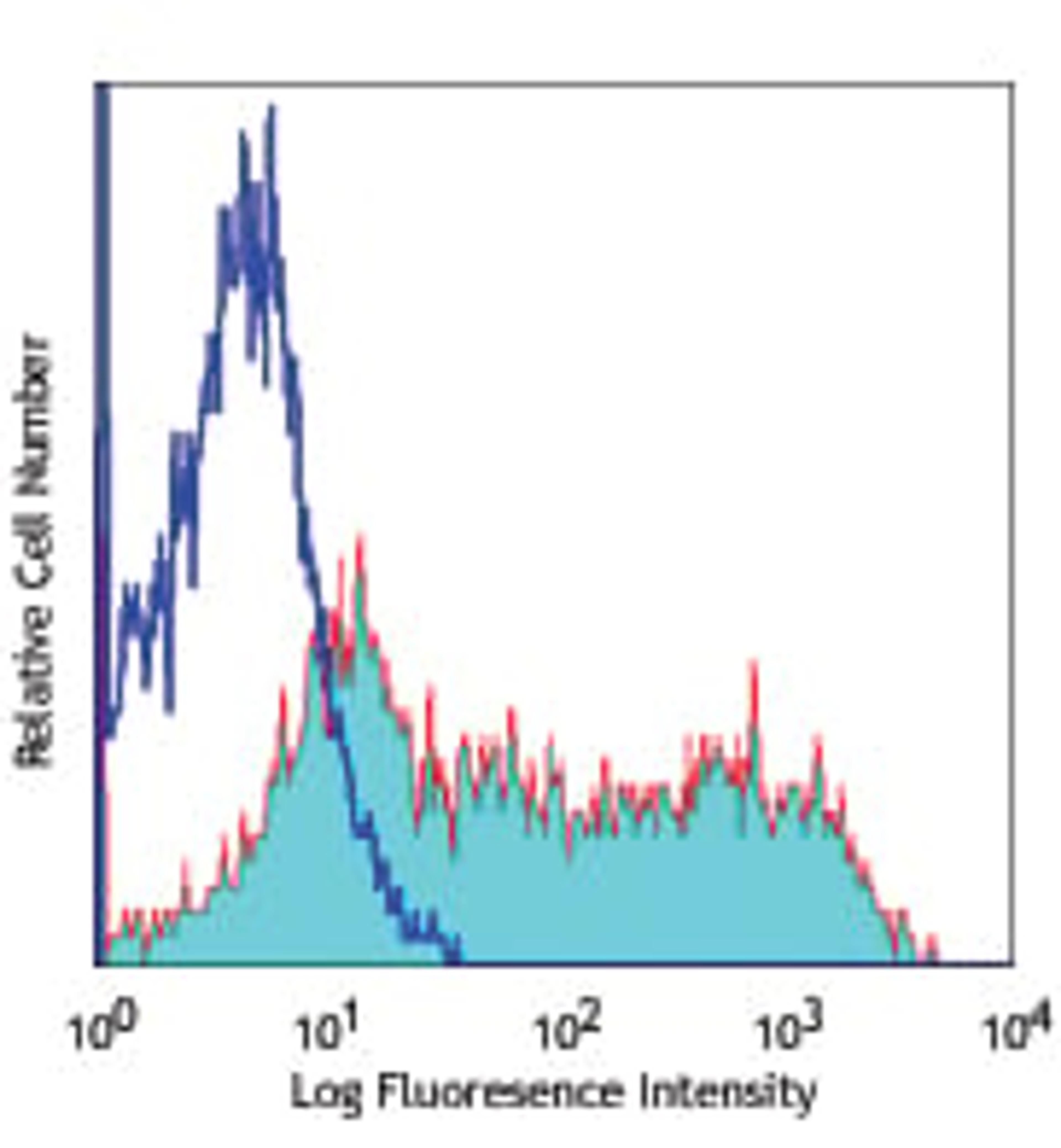
![Flow Cytometry: beta-Actin Antibody (AC-15) [NB600-501] - Analysis using the HRP conjugate of NB600-501. Electropherogram image(s) of corresponding Simple Western lane view. Beta-Actin antibody was used at 1:500 dilution on Hela, MCF-7, SH-SY5Y, & Jurkat lysate(s).](https://cdn.sanity.io/images/f5b6mtfn/antibodies-prod/abc36365e4291dc306bd1a8e1d212c04af18010f-400x250.jpg?w=3840&q=75&fit=clip&auto=format)
beta-Actin Antibody (AC-15)
- Manufacturer
- Novus Biologicals, LLC
- Catalog Number
- NB600-501
- Host
- Mouse
- Reactivity
- Bovine, Human, Mouse, Pig (Swine)
- Applications
- ELISA, Flow Cytometry, Immunocytochemistry, Immunohistochemistry, Immunohistochemistry (Frozen Sections), Immunohistochemistry (Paraffin-Embedded Sections), Immunoprecipitation, Western Blotting
![Immunocytochemistry/Immunofluorescence: HIF-1 alpha Antibody [NB100-479] - Detection of HIF-1 Alpha (green) in RCC4 cells using NB100-479. Nuclei (Blue) were counterstained using Hoechst 33258.](https://cdn.sanity.io/images/f5b6mtfn/antibodies-prod/5512a02e1c1e276d7261b292580c94dfd91a17c7-400x96.jpg?w=3840&q=75&fit=clip&auto=format)
![Immunocytochemistry/Immunofluorescence: LC3/MAP1LC3A Antibody [NB100-2331] - LC3 antibody was tested in HeLa cells with Dylight 488 (green). Nuclei and alpha-tubulin were counterstained with DAPI (blue) and Dylight 550 (red).](https://cdn.sanity.io/images/f5b6mtfn/antibodies-prod/0196282f2adb49bb283ea442ece1bb30f88a5885-400x300.jpg?w=3840&q=75&fit=clip&auto=format)
![Immunocytochemistry/Immunofluorescence: APE Antibody (13B8E5C2) [NB100-116] - Immunocytochemical detection of APE-ref-1 in breast cancer cell line MDS231.](https://cdn.sanity.io/images/f5b6mtfn/antibodies-prod/8b1c32229e7686b3db8bf9620197c949b433fb11-400x300.jpg?w=3840&q=75&fit=clip&auto=format)
![Flow Cytometry: xCT Antibody [NB300-318] - xCT antibody was tested at 1:400 in HepG2 cells using an Alexa Fluor 488 secondary (shown in purple). M1 is defined by unstained cells.](https://cdn.sanity.io/images/f5b6mtfn/antibodies-prod/b24c08cace855ed0db4d7ed0b41aac61229de0cb-400x307.jpg?w=3840&q=75&fit=clip&auto=format)
![Western Blot: active/cleaved Caspase 8 Antibody [NB100-56116] - analysis of Caspase 8 in NRK whole cell lysate using anti-active/cleaved Caspase 8 antibody. Image from verified customer review.](https://cdn.sanity.io/images/f5b6mtfn/antibodies-prod/f28b8f68166461c9ead094c3d9a2e5640d3875e5-237x400.jpg?w=3840&q=75&fit=clip&auto=format)
![Immunocytochemistry/Immunofluorescence: DNMT1 Antibody [NB100-264] - The Dnmt1 antibody was tested in HeLa cells at a 1:50 dilution against Dylight 488 (Green). Alpha-tubulin and nuclei were counterstained against Dylight 550 (Red) and DAPI (Blue), respectively.](https://cdn.sanity.io/images/f5b6mtfn/antibodies-prod/d53959ff27112bb4036b9d5a89b58b070b341496-400x300.jpg?w=3840&q=75&fit=clip&auto=format)
![Immunohistochemistry-Paraffin: iNOS Antibody [NB300-605] - Both normal and cancer biopsies of deparaffinized human Heart tissue.](https://cdn.sanity.io/images/f5b6mtfn/antibodies-prod/890af3ce58f15c655c7d5a73cdc4686517d9e191-400x200.jpg?w=3840&q=75&fit=clip&auto=format)
![Immunocytochemistry/Immunofluorescence: Fibrillarin Antibody (38F3) [NB300-269] - Human SH-SY5Y cells stained with NB300-269, showing prominent specular nucleolar staining. The nuclei are counter stained with DAPI (blue), so these spots appear very pale blue. Neurofilament heavy protein was stained with NB300-217 (red).](https://cdn.sanity.io/images/f5b6mtfn/antibodies-prod/856d433a1206f6a5f24493c28805f673fd9c6dbf-400x314.jpg?w=3840&q=75&fit=clip&auto=format)
![Flow Cytometry: Tenascin C Antibody (4C8MS) [NB110-68136] - Intracellular flow cytometric staining of 1 x 10^6 MCF-7 cells using Tenascin C antibody (dark blue). Isotype control shown in orange. An antibody concentration of 1 ug/1x10^6 cells was used.](https://cdn.sanity.io/images/f5b6mtfn/antibodies-prod/8b6c8c73df8fd02df534c469d1865eb90b75e422-400x391.jpg?w=3840&q=75&fit=clip&auto=format)
![Immunocytochemistry/Immunofluorescence: PKC alpha Antibody (MC5) [NB600-201] - PKC alpha (MC5) antibody was tested in SH-SY5Y cells with Dylight 488 (green). Nuclei and beta-tubulin were counterstained with DAPI (blue) and Dylight 550 (red).](https://cdn.sanity.io/images/f5b6mtfn/antibodies-prod/6de4ab391081c8a64552ddd3b98289b890f02a32-400x386.jpg?w=3840&q=75&fit=clip&auto=format)
![Western Blot: AG-2 Antibody [NBP2-27393] - Analysis using the Azide Free version of NBP2-27393. Detection of AGR2 using AGR2 antibody. MCF7 (A), Caco-2 (B) and human stomach lysate (C) probed with AGR2 antibody at 1:100. were used for this test. The higher molecular weight band of variable intensity at ~30 kDa is uncharacterized and may represent a form of AGR2.](https://cdn.sanity.io/images/f5b6mtfn/antibodies-prod/fcf1ae52b79670155bef2c758325d195959983e8-243x400.jpg?w=3840&q=75&fit=clip&auto=format)
![Immunohistochemistry-Paraffin: Hsp47 Antibody (M16.10A1) [NBP1-97491] - Analysis of paraffin-embedded tissue section of rat carotid artery 14 days after balloon-injury to the artery, stained using Hsp47 (Colligin) mAb (M16.10A1).](https://cdn.sanity.io/images/f5b6mtfn/antibodies-prod/79f1700eca231a25bc4a07de2a53ac9214bb795f-400x279.jpg?w=3840&q=75&fit=clip&auto=format)
![Immunohistochemistry-Paraffin: eNOS Antibody [NB300-500] - Staining of eNOS in human lung tissue.](https://cdn.sanity.io/images/f5b6mtfn/antibodies-prod/f74438569f05b62eada2a2b504d48317a5881a50-400x352.jpg?w=3840&q=75&fit=clip&auto=format)
![Immunohistochemistry-Paraffin: GFAP Antibody (GA5) [NBP2-29415] - Formalin-paraffin human brain stained with GFAP Ab (GA-5). Note cytoplasmic staining.](https://cdn.sanity.io/images/f5b6mtfn/antibodies-prod/d6bda0c41e0766e2c4f7be648cfb5dbe507259b6-400x300.jpg?w=3840&q=75&fit=clip&auto=format)
![ELISA: Twist-1 Antibody (10E4E6) [NBP2-37364] - Red: Control Antigen (100ng); Purple: Antigen (10ng); Green: Antigen (50ng); Blue: Antigen (100ng);](https://cdn.sanity.io/images/f5b6mtfn/antibodies-prod/508ca5d7f6aee3e7c331507ff124f7863faa9651-400x333.jpg?w=3840&q=75&fit=clip&auto=format)
![Flow (Cell Surface): CD163 Antibody (6E10.1G6) [NBP2-36494] - Human peripheral blood mononuclear cells / PBMCs were tested in FLOW with 2.5ug / million cells of CD163 antibody clone 6E10.1G6 and 0.5ug/million cells of goat anti-Mouse IgG-PE secondary antibody. Cells alone (Blue), Isotype control IgG2b kappa (Green) and CD163 antibody clone 6E10.1G6 (Red).](https://cdn.sanity.io/images/f5b6mtfn/antibodies-prod/652ffed3356077f1515168a3ba24b0b8926a947f-400x329.jpg?w=3840&q=75&fit=clip&auto=format)
![Immunocytochemistry/Immunofluorescence: GATA-2 Antibody [NBP1-82581] - Staining of human cell line U-251 MG shows positivity in nucleus but excluded from the nucleoli. Antibody staining is shown in green.](https://cdn.sanity.io/images/f5b6mtfn/antibodies-prod/6fb2493a611426f124d92e618a25d4ea3c37eee8-376x400.jpg?w=3840&q=75&fit=clip&auto=format)

![Immunohistochemistry-Paraffin: ErbB2/HER2 Antibody (SP3) [NBP1-49793] - c-erbB-2/HER-2](https://cdn.sanity.io/images/f5b6mtfn/antibodies-prod/a96dcd257e83e783c4f6d61a79b542053c8980d9-395x400.jpg?w=3840&q=75&fit=clip&auto=format)
![Western Blot: IL17RA Antibody (49M4D2) [NBP2-25258] - analysis of an IL-17RA recombinant protein fragment and human heart lysate using IL-17RA antibody at 0.5 and 3 ug/ml, respectively. goat anti-mouse Ig HRP secondary antibody and PicoTect ECL substrate solution were used for this test.](https://cdn.sanity.io/images/f5b6mtfn/antibodies-prod/9f408401a12f2eac8b3b79740d4429d35a61b50a-172x400.jpg?w=3840&q=75&fit=clip&auto=format)
![Immunocytochemistry/Immunofluorescence: Substance P Antibody [NB300-187] - Detection of Substance P in rat spinal cord dorsal horn (red fluorescence). DAPI (blue) was used as counter stain.](https://cdn.sanity.io/images/f5b6mtfn/antibodies-prod/f0425c6c586c0d2f1b8a05c2d1062775ef172dd2-400x316.jpg?w=3840&q=75&fit=clip&auto=format)
![Flow Cytometry: EIF2S1 Antibody (3H4) [NBP2-02669] - HEK293T cells transfected with either overexpression plasmid (Red) or empty vector control plasmid (Blue) were immunostained by anti-eIF2A antibody, and then analyzed by flow cytometry.](https://cdn.sanity.io/images/f5b6mtfn/antibodies-prod/abd3273dd9ebaa14f66cf3419e05a05b8d4c588c-400x265.jpg?w=3840&q=75&fit=clip&auto=format)
![Western Blot: DNA Ligase III Antibody (1F3) [NBP1-41190] - Sample (30 ug of whole cell lysate) A: Hela 7. 5% SDS PAGE, antibody diluted at 1:500.](https://cdn.sanity.io/images/f5b6mtfn/antibodies-prod/6f8edcf37431f0f5d4557704060236361be4e3a1-225x400.jpg?w=3840&q=75&fit=clip&auto=format)