TIA-1 Antibody (G-3)
Product Details
- Cat. No.
- sc-166247
- Type
- Primary Antibody
- Clonality
- Monoclonal
- Host
- Mouse
The supplier does not provide quotations for this antibody through SelectScience. You can search for similar antibodies in our Antibody Directory.
Description
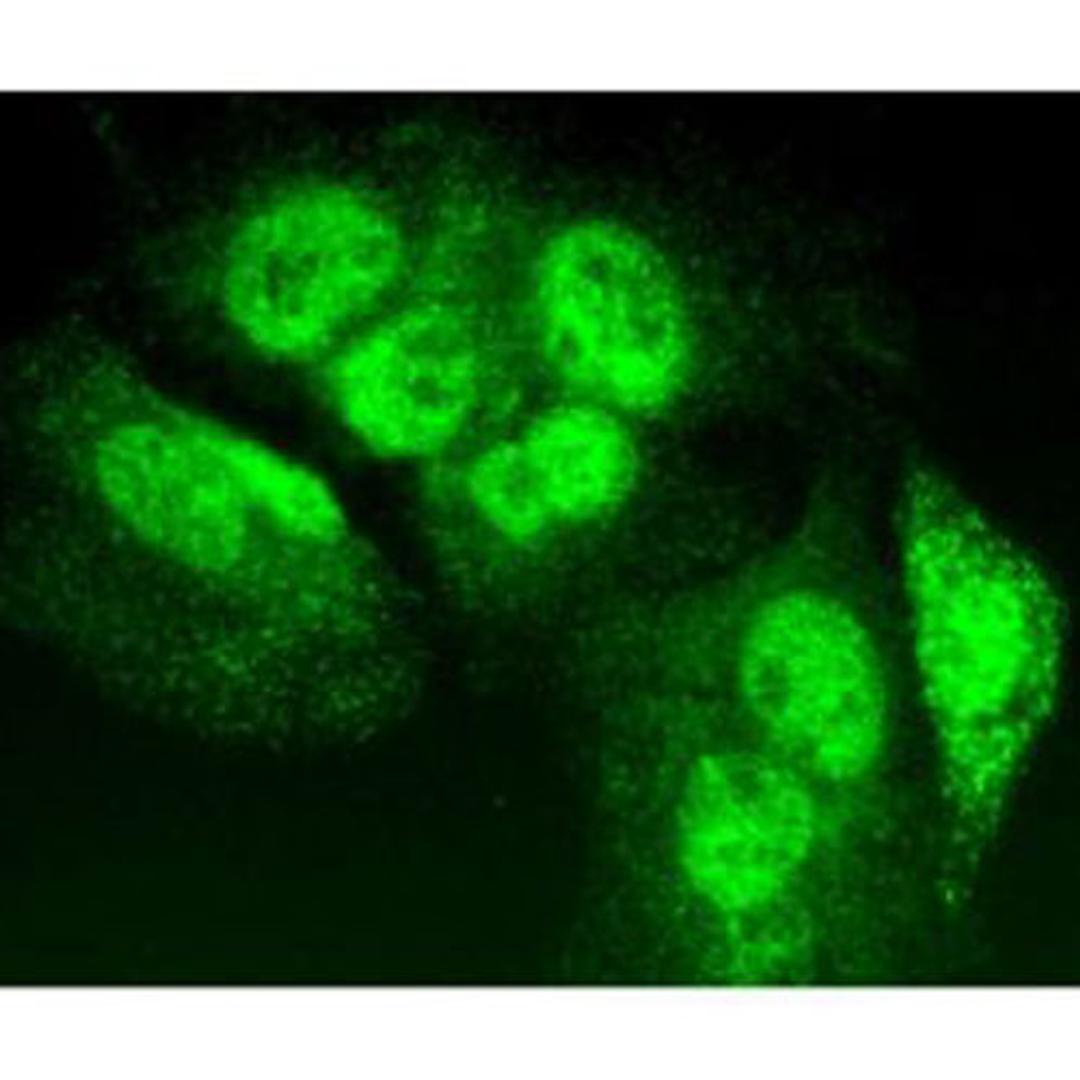
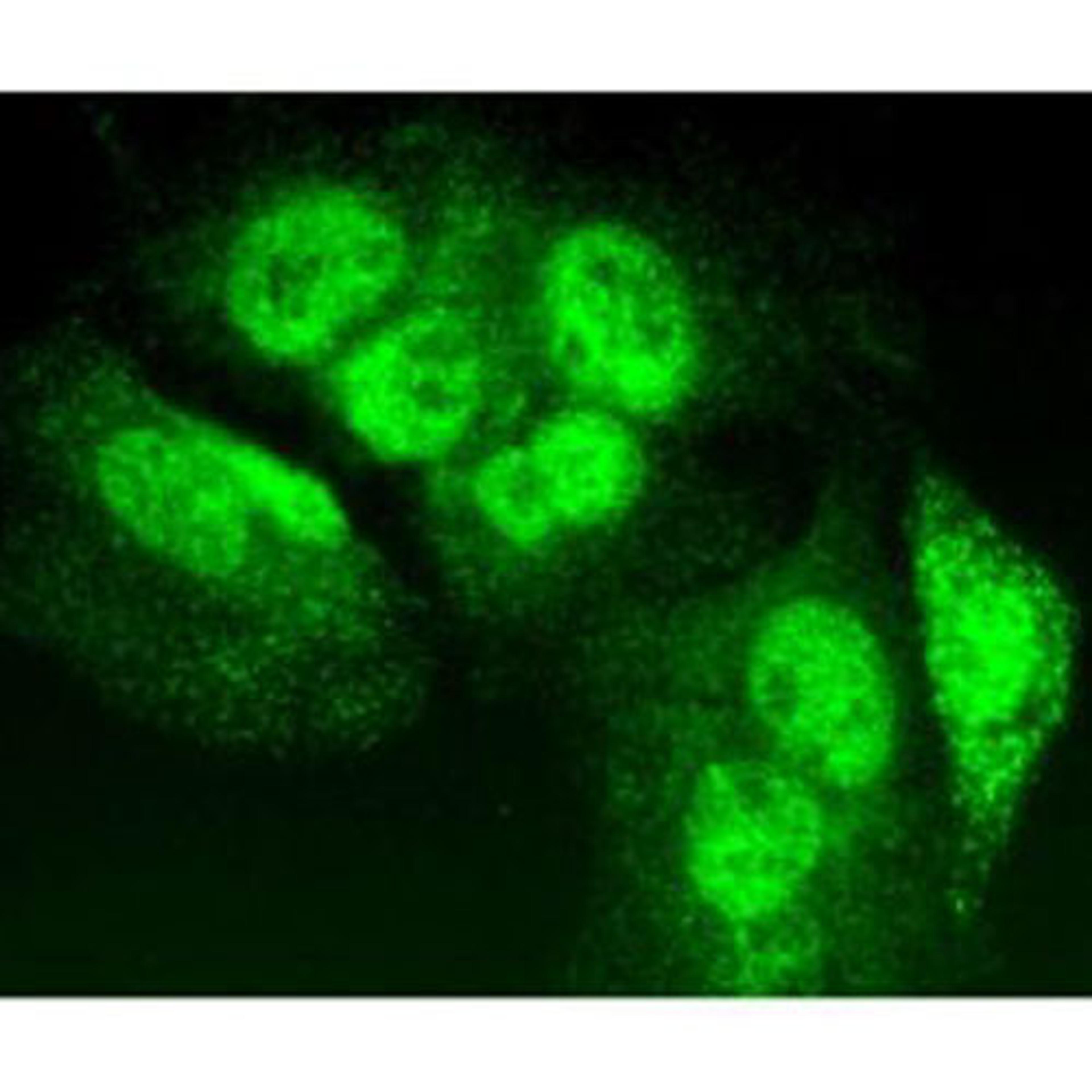
TIA-1 Antibody (G-3) is a high quality monoclonal TIA-1 antibody (also designated TIA1 antibody, Nucleolysin TIA-1 antibody or p40-TIA-1 antibody) suitable for the detection of the TIA-1 protein of mouse, rat and human origin. TIA-1 Antibody (G-3) is available as both the non-conjugated anti-TIA-1 antibody form, as well as multiple conjugated forms of anti-TIA-1 antibody, including agarose, HRP, PE, FITC and multiple Alexa Fluor® conjugates. FAS, also referred to as CD95 or APO-1, is a type I transmembrane protein that plays a central role mediating viral immunity. TIA-1 and TIAR are two closely related proteins that possess three RRMs (RNA recognition motifs), designated RRM 1, 2 and 3. Although both TIA-1 and TIAR are thought to function as mediators of apoptotic cell death, their specific roles in such pathways are unknown. Unlike TIA-1, which is found in the granules of cytotoxic lymphocytes, TIAR expression is limited to the nucleus and found in a much broader range of cells including, but not limited to, cells of hematopoietic origin. TIAR is translocated to the cytoplasm shortly after FAS ligation and this event immediately proceeds the onset of DNA fragmentation. A novel serine/threonine kinase that is activated as a result of FAS ligation, designated FAST (FAS-activated serine/threonine), shows kinase specificity towards both TIA-1 and TIAR. In unstimulated Jurkat cells, FAST resides in the cytoplasm as a highly phosphorylated protein and is quickly dephosphorylated and activated in response to stimulated FAS.
Biological Information
- Clonality: Monoclonal
- Host: Mouse
- Reactivity: Human, Mouse, Rat
Handling
- Quantity: 200 µg/ml
- Specificity: 1
Applications
- ELISA (ELISA)
- Immunofluorescence (IF)
- Immunohistochemistry (Paraffin-Embedded Sections) (IHC (P))
- Immunoprecipitation (IP)
- Western Blotting (WB)
It does not work on brain tissue.
Western blot
We bought this antibody to detect endogenous protein in Human brain tissue by western blot. There is no signal at all loading up to 40ug of protein and using the antibody 1:500. A test on rat brain showed the same results. Unfortunately not useful on brain tissues at all.
Review Date: 20 Feb 2021 | Santa Cruz Biotechnology Inc.


