3D Electron Microscopy for Life Sciences
Special Issue of the Journal of Microscopy celebrates the future of cell imaging
21 Apr 2016

For the past 50 or so years, the ability to obtain extensive three-dimensional information from cells has been the preserve of those brave electron microscopists willing to undertake extensive serial thin sectioning followed by manual or semiautomated reconstruction. More recently, the technique of transmission electron tomography has enabled the high-resolution study of subcellular structure at extremely high resolution in 3-D. However, to obtain reconstructions to any great depth using this technique is time-intensive, involves collecting serial tomograms, is extremely skillful and reconstructions can take many weeks to complete.
The development of high-resolution field emission scanning electron microscopes (FE-SEM) has enabled biologists to explore the use of the SEM to study cell ultrastructure in resin-embedded material using traditional TEM preparative techniques. Combined with ion beam milling (FIB-SEM) techniques or the ability to undertake ultramicrotomy within the SEM specimen chamber, known as serial block face imaging (SBFSEM, SB-EM), it is now possible to collect data sets of many microns in the z-axis and reconstruct organelle organization in whole cells or tissues.
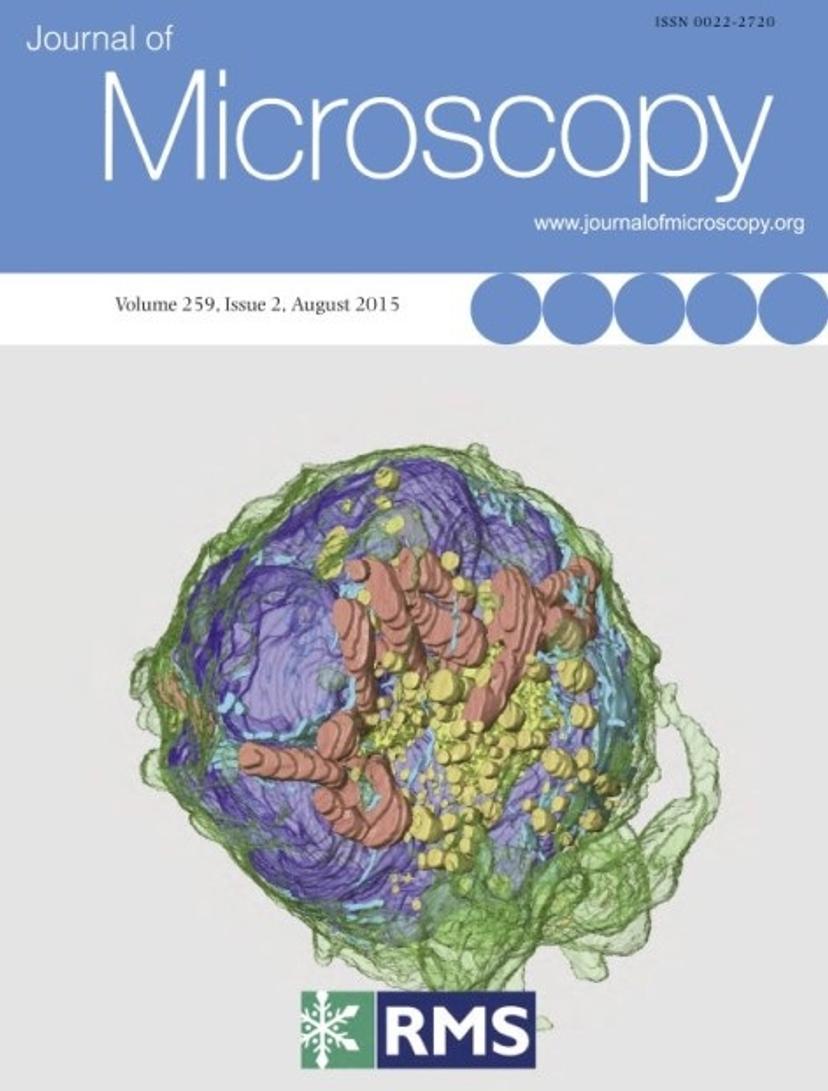
The August 2015 Issue of the Journal of Microscopy, edited jointly by Chris Hawes (Oxford Brookes University) and Eric Hummel (ZEISS Microscopy) contains a series of papers exploring the developments and applications of the use of scanning electron microscopy to study cell ultrastructure. This includes papers on correlative fluorescence and SEM imaging, SBFSEM and FIB-SEM, through to array tomography of sections imaged by SEM and high-throughput imaging using a multi-beam SEM.
ZEISS together with the RMS and the Journal of Microscopy jointly present the open access articles from the August 2015 issue.