8 Recent Advances in Bio Imaging and Microscopy
Discover the latest products and methods for the imaging of biological samples
31 Aug 2015
Discover the latest methods and products for imaging biological samples
1. APPLICATION NOTE: Multiple Images from a Single Tissue Section with Dual Polarity Desorption Electrospray Ionization MS
Learn how DESI imaging using Waters' SYNAPT® G2-Si HDMS Mass Spectrometer enables sequential analysis of a single tissue section for comprehensive molecular profiling. Mass spec imaging of tissue sections was accomplished using a SYNAPT® G2-Si HDMS Mass Spectrometer equipped with a 2D-DESI source, and data was generated and analyzed using HDI Software v1.3. Download method.
2. PRODUCT: Solutions for the Visualization and Analysis of Big Image Data in Life Sciences
New microscopy technologies, such as light sheet fluorescence microscopy (LSFM) or clearing methods, allow the imaging of large samples at high resolution or high frame rates. ZEISS has teamed up with arivis AG to offer complete solutions, from initial image acquisition to final results, to overcome common challenges in handling, processing, and analyzing multi-terabyte data sets. Learn more.
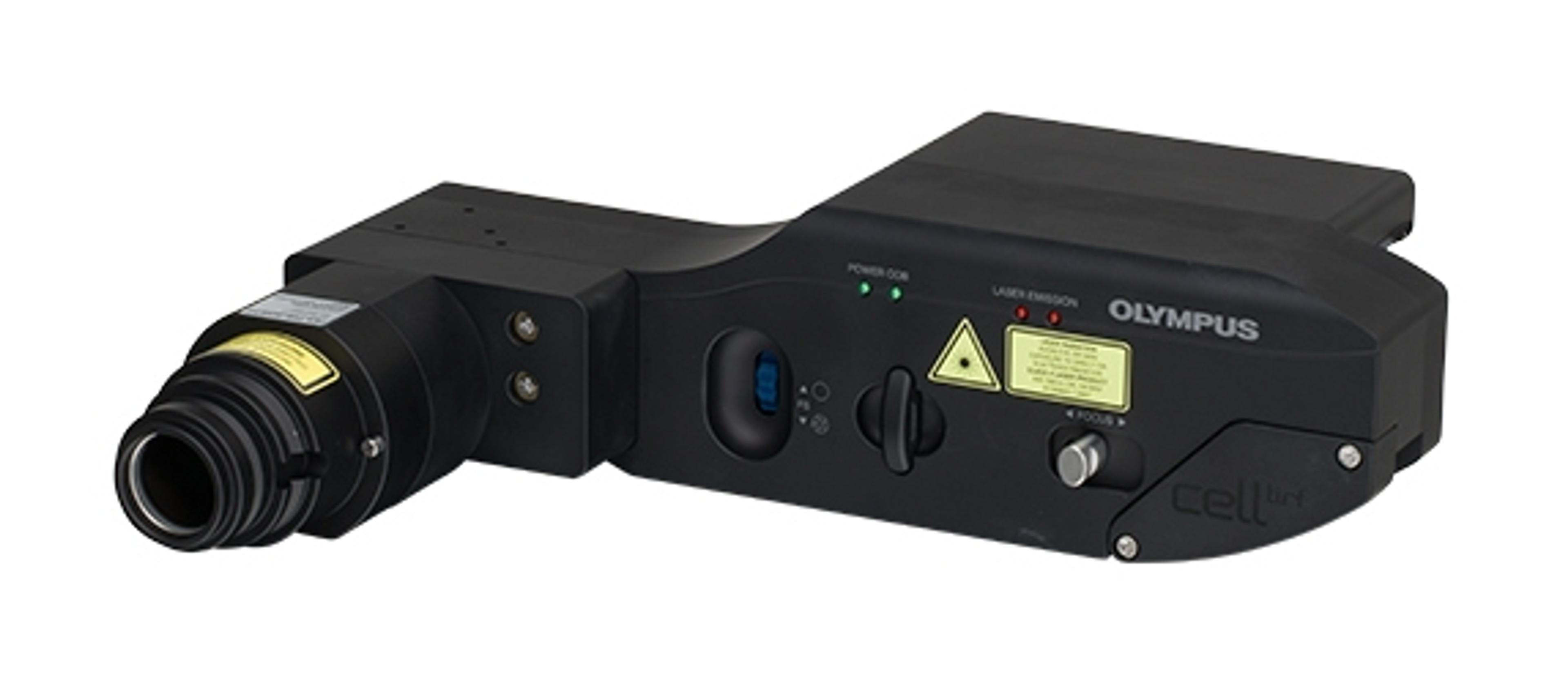
3. PRODUCT: Unmatched Optics, Laser Control and Accuracy from Expanded Olympus cellTIRF Family
The recently expanded Olympus cellTIRF family represents the latest technology in advanced TIRF solutions, providing a series of features for precise imaging with effortless control. By matching quality optics with high-end motorized TIRF devices, Olympus offers a system that enables easy access to super-resolution microscopy, performed using single-molecule localization techniques. Read more.
4. WEBINAR: Quantification of NF-ĸB Signaling in Living Cells using the Operetta High Content Imaging System
Discover how PerkinElmer’s Operetta® High Content Imaging System can be used to study the dynamics of the NF-ĸB family of transcription factors, pivotal in immunity and cell response, in living cells. NF-ĸB signaling is an attractive target in the development of drugs against inflammation-related diseases. Watch on-demand.
5. APPLICATION NOTE: Lumenera’s Scientific-Grade Cameras Offer Imaging Solutions for Growing Digital Pathology Market
Pathology is intricately connected to all medical advancements, as the study and diagnosis of disease through examination of organs, tissues, and bodily fluids. While microscopes still hold a central role in this science, digital pathology stands as an equally critical evolution in this field. Digital pathology is the practice of digitizing glass slides and managing the resultant information for later educational, diagnostic, and analytic purposes. In this application note, the use of Lumenera’s scientific grade cameras in digital pathology applications is explored. Download method.
6. VIDEO: Improved Imaging of Large Cleared Samples with ZEISS Lightsheet Z.1
Tissue clearing allows you to image deep into large biological samples such as tissue sections, brains, embryos, organs, spheroids or biopsies. Light sheet fluorescence microscopy (LSFM) enables imaging of cleared specimens with exceptional light efficiency, speed and next to no photo damage. Watch this video to learn how to mount cleared samples in ZEISS Lightsheet Z.1. Watch video.
7. ARTICLE: My Lab Essentials: Why UV isn't the Enemy
In the latest interview in our Lab Essentials series, Dr Richard Weller, Senior Lecturer in Dermatology at the University of Edinburgh, explains the effect of sunlight on the cardiovascular system, how we could be doing more harm than good by avoiding the sun in the name of health, and how microscopy is essential in his research. Read more.
8. BUYING GUIDE: Download the New Microscopy Buying Guide Now!
New microscopy techniques are continually developing as solutions to imaging problems. If you are looking to invest in microscopy equipment, this essential SelectScience Buying Guide provides you with all the information you need to help you to make the right decision, based on your requirements. Download now.

