Advances in biomarker diagnostics for Alzheimer’s disease
Revvity’s Euroimmun shares insights on the role of amyloid-beta, tau, neurofilament and APOE genotype in Alzheimer’s disease diagnostics
12 Aug 2025
The need for early and accurate diagnosis of Alzheimer’s disease (AD) has become more pressing with the clinical availability of disease-modifying therapies. These agents, which aim to slow cognitive decline by targeting amyloid pathology, are most effective when administered in the early stages of mild cognitive impairment. Moreover, etiologic diagnosis is essential as an inclusion criterion for clinical trials of new therapies.
Current frameworks define AD by its biology rather than clinical presentation alone. Biomarker evidence of AD neuropathological changes is now sufficient to establish a diagnosis in cognitively impaired individuals. In unimpaired persons, abnormal biomarker results are associated with an elevated risk of progression to symptomatic disease.1
Core diagnostic biomarkers
The neuropathological hallmarks of AD are extracellular amyloid-beta (Aβ) plaques and intracellular neurofibrillary tangles of hyperphosphorylated tau protein. The core biomarkers for AD map onto the amyloid-beta or tauopathy pathways.
Aβ and tau biomarkers are detected by positron emission tomography (PET) or biofluid measurements. New criteria from the Alzheimer’s Association propose the following can be diagnostic for AD in symptomatic persons: amyloid PET or a hybrid ratio of Aβ 42/40, p-tau 181/Aβ 42 or t-tau/Aβ 42 determined in CSF. Plasma assays are only acceptable if they have an accuracy equivalent to CSF assays in the intended-use population.1, 2
Neurofilament markers
Non-specific biomarkers are those that are important in the AD pathogenic pathway but are also involved in other brain diseases. For example, neurofilament light (NfL) is a sensitive indicator of large-caliber axonal injury and an upcoming biomarker for neurodegenerative disease diagnostics. Elevated levels of NfL can occur in AD as well as in multiple sclerosis, amyotrophic lateral sclerosis (ALS) and traumatic brain injury.1 Of note, the phosphorylated heavy chain of neurofilament (pNfH) also serves as an indicator of axonal damage and is considered the most sensitive diagnostic marker for ALS.3 Some further promising AD biomarkers such as glial fibrillary acidic protein (GFAP) are involved in inflammatory and immune processes.
Detection methods
Although PET is considered the reference standard for detecting abnormal amyloid, biofluid analytics may be preferred for routine diagnostics due to lower costs and the option to perform multiple biomarker tests at the same time.
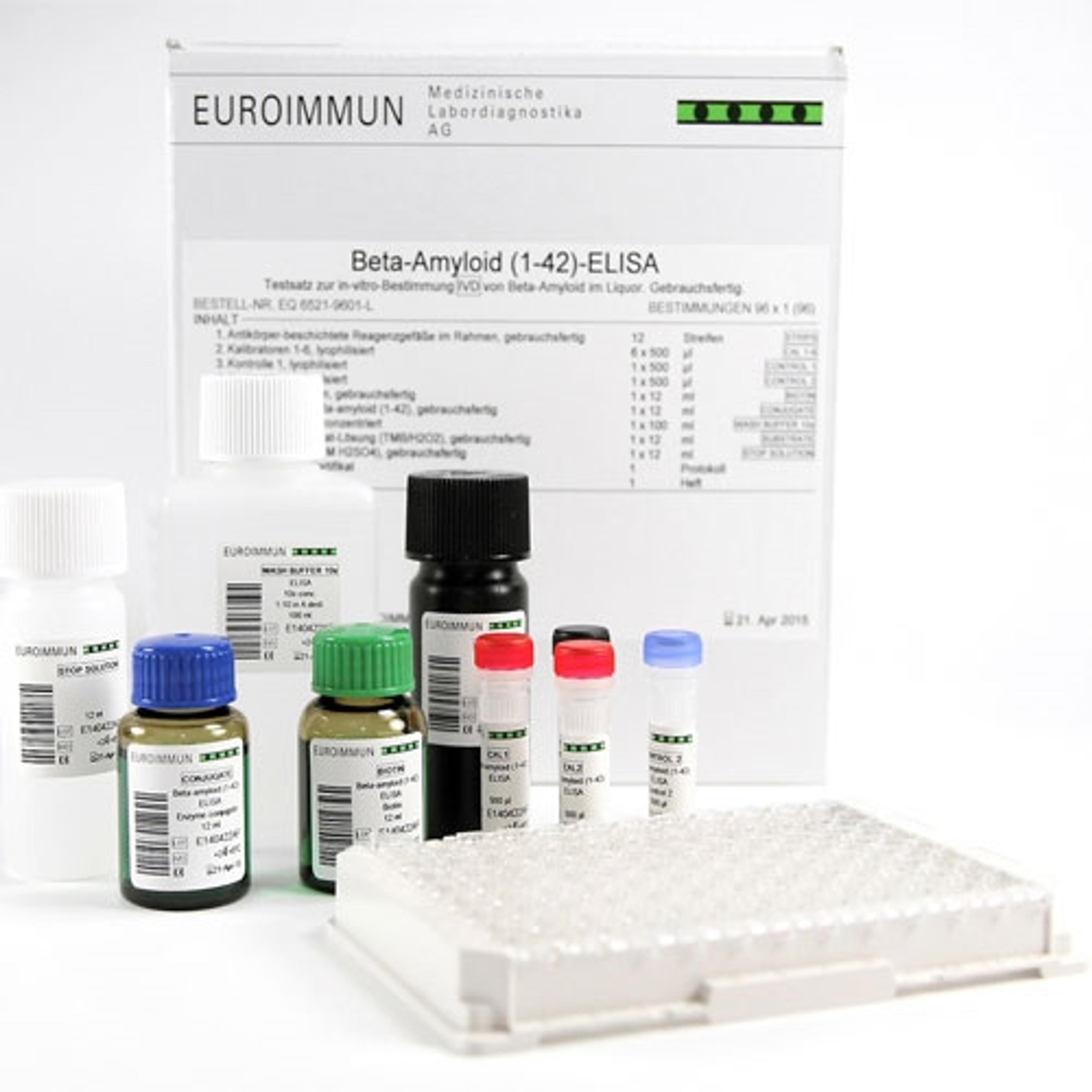
Chemiluminescence immunoassay (ChLIA) and ELISA are among the most common methods used for biofluid measurements. While both techniques offer robust measurements, ChLIAs are increasingly favoured by laboratories due to their higher sensitivity and faster turnaround times.
A panel of fully automated ChLIAs for measurement of Aβ 42/40, t-tau and p-tau181 in CSF (Euroimmun) demonstrated reliable analytical and clinical performance in published collaborative evaluation studies, confirming their suitability to support the differential diagnosis of AD from similar neuropsychiatric disorders.4, 5 The corresponding ELISA range covers the same core parameters and is complemented by a pNf-H ELISA and an NfL ELISA (available soon) for extended biomarker profiling. Ongoing development of the assay portfolio is focused on additional neurodegeneration markers as well as robust blood-based assays.
CSF versus blood
While blood-based biomarker (BBM) tests are gaining momentum, their performance varies widely, and their clinical interpretation across varying patient cohorts is not well standardised. Comorbidities such as renal insufficiency, obesity and cardiovascular disease may also influence levels of BBMs. Notably, plasma Aβ 42/40 shows a smaller change with amyloid positivity than CSF, resulting in worse clinical robustness. p-tau217 is a more promising plasma marker correlating with AD neuropathological changes in clinical studies.1, 2, 6
The recently updated German S3 Guideline on Dementias advises against using BBMs as standalone tests in practice, recommending that BBM findings be confirmed with established biomarker modalities.6
Nevertheless, BBMs are anticipated to play a central role in future diagnostic pathways due to their accessibility, lower invasiveness and scalability. Minimum performance standards for clinical use of BBM tests have recently been defined to guide the development and evaluation of new assays.2
APOE genetic risk factor
An important consideration when implementing anti-beta-amyloid therapies is the potential for serious or life-threatening side effects in some individuals. These include amyloid-related imaging abnormalities (ARIA) with edema and/or effusion (ARIA-E) or hemorrhagic changes (ARIA-H). The risk of ARIA is closely linked to the apolipoprotein E (APOE) genotype, with homozygous carriers of the ε4 allele at highest risk.
APOE genotyping is therefore recommended for risk stratification prior to initiating anti-amyloid therapies.7 In countries that restrict treatment with anti-amyloid therapies to ε4 noncarriers and heterozygotes, an APOE genetic test is a requisite prior to treatment.
The APOE genotype can be easily determined by real-time PCR (EURORealTime APOE, Euroimmun), which enables precise typing of the six possible genotypes ε2/ε2, ε2/ε3, ε2/ε4, ε3/ε3, ε3/ε4 and ε4/ε4. The analysis supports proactive evaluation of the individual ARIA risk and a more personalised approach to therapy.
Regulatory status, precise intended use and product availability for the mentioned Euroimmun tests must be verified for the user‘s individual jurisdiction.
References
1. Jack CR Jr, Andrews JS, Beach TG et al. Revised criteria for diagnosis and staging of Alzheimer's disease: Alzheimer's Association Workgroup. Alzheimers Dement. 2024 Aug;20(8):5143-5169. doi: 10.1002/alz.13859.
2. Schindler SE, Galasko D, Pereira AC et al. Acceptable performance of blood biomarker tests of amyloid pathology — recommendations from the Global CEO Initiative on Alzheimer’s Disease. 2024; Nat Rev Neurol 20, 426-439. doi.org/10.1038/s41582-024-00977-5
3. Heckler I, Venkataraman I. Phosphorylated neurofilament heavy chain: a potential diagnostic biomarker in amyotrophic lateral sclerosis. J Neurophysiol. 2022 Mar 1;127(3):737-745. doi: 10.1152/jn.00398.2021.
4. Römpler K, Arendt P, Brix B et al. Evaluation of the EUROIMMUN automated chemiluminescence immunoassays for measurement of four core biomarkers for Alzheimer’s disease in cerebrospinal fluid. Practical Laboratory Medicine. 2024 Aug; Vol 41. doi.org/10.1016/j.plabm.2024.e00425
5. Arendt P, Römpler K, Brix B et al. Differentiation of Alzheimer's disease from other neurodegenerative disorders using chemiluminescence immunoassays measuring cerebrospinal fluid biomarkers. Frontiers in Dementia. Section Genetics and Biomarkers of Dementia. 2024; Vol 3. doi: 10.3389/frdem.2024.1455619
6. S3 Guideline: Dementias - Living Guideline (Version 5.2, Feb 28, 2025). German Association for Psychiatry, Psychotherapy and Psychosomatics (DGPPN), German Society of Neurology (DGN). AWMF Reg-Nr. 038-013. https://register.awmf.org/de/leitlinien/detail/038-013
7. Cummings J et al. Lecanemab: Appropriate use Recommendations. J Prev Alzheimer’s Dis. 2023;10(3):362-377. doi: 10.14283/jpad.2023.30.