Bio-Rad’s New Workflow Enables Highly Sensitive Evaluation of Antibodies Used to Measure HCP Impurities in Biotherapeutics
28 Jan 2016Bio-Rad Laboratories, Inc. today announced the launch of a new simplified and robust workflow to more effectively evaluate antibodies used for measuring residual host cell protein (HCP) impurities in biotherapeutics. The two-dimensional electrophoresis (2-DE)- and western blotting (WB)-based workflow will be unveiled during the Well Characterized Biotechnology Pharmaceutical (WCBP) Symposium in Washington, D.C. January 29–31.
Regulatory agencies worldwide including the U.S. Food and Drug Administration (FDA) require that residual HCP impurities in biotherapeutics remain below levels detectable using a highly sensitive analytical method (PTC 1997). Failure to meet this requirement puts patients at risk, an issue recently documented by a biopharmaceutical company forced to put a clinical trial on hold when patients developed a positive response to its product’s HCP.
“It can take a company several months and tens of thousands of dollars to develop an adequately immunospecific host cell protein reagent, so there is a lot time and money riding on the validity of that reagent,” said Nadine Ritter, senior CMC consultant at the Biologics Consulting Group.
The most widely used quality control method for determining the presence of HCPs is the ELISA, a test that uses a polyclonal anti-HCP antibody mixture raised against HCPs. To ensure the accuracy of HCP ELISA results, ICH Q6B guidelines state that polyclonal antibodies must be “capable of detecting a wide range of protein impurities.” The standard workflow for evaluating this range — defined as the percentage of HCPs that are detectable by polyclonal antibodies — involves 2-DE separation of host cell protein preparations, then transfer of the proteins from the gel to a solid support membrane, followed by western blotting with the polyclonal anti-HCP antibodies.
Although this workflow is widely used, several key issues remain, including time-intensive optimization of the components of 2-DE separation (IEF and SDS-PAGE) and variable protein transfer efficiencies. These issues reduce the overall reliability of 2-DE and western blotting results and thus lower confidence in the evaluation of anti-HCP antibodies. By integrating unique instruments and consumables into a workflow solution for addressing these issues, Bio-Rad offers highly confident evaluation of the range of protein impurities detectable by anti-HCP antibodies.
Results Presented at WCBP Demonstrate Workflow’s Effectiveness
Bio-Rad researchers will present results at WCBP that demonstrate the effectiveness of their 2-DE/WB-based workflow for evaluating polyclonal anti-HCP antisera. Bio-Rad senior staff scientist Tom Berkelman and his colleagues generated optimized host cell protein separations via 2-DE and integrated them with rapid semi-dry blotting, fluorescent protein staining, and western blotting to confidently evaluate the antisera within two days.
“Bio-Rad’s 2-DE/WB-based technique allows the detection of HCP impurities often unseen in one-dimensional electrophoresis,” said Karin Abarca Heidemann, PhD, R&D director at antibody development firm, Rockland Immunochemicals, Inc. “This is a significant benefit in the development of our HCP antibodies, as two-dimensional electrophoresis delivers improved insights into the quality and immunocoverage of antibodies against host cell contamination. Rockland is collaborating successfully with Bio-Rad on developing an improved standardized immunoassay for HCP detection using its 2-DE/WB workflow, the result of which will be presented during the WCBP Symposium.”
Bio-Rad’s New Workflow
Bio-Rad’s HCP antibody evaluation workflow consists of the following steps and takes two days to complete:
1. Separate proteins based on isoelectric point (pI) in IPG strips using the PROTEAN® i12™ IEF system (the first dimension of 2-DE). The individual lane control feature — unique to the PROTEAN i12 system — enables fast optimization of isoelectric focusing (IEF) conditions and easy application of different optimized conditions for highly reproducible, simultaneous IEF of multiple samples.
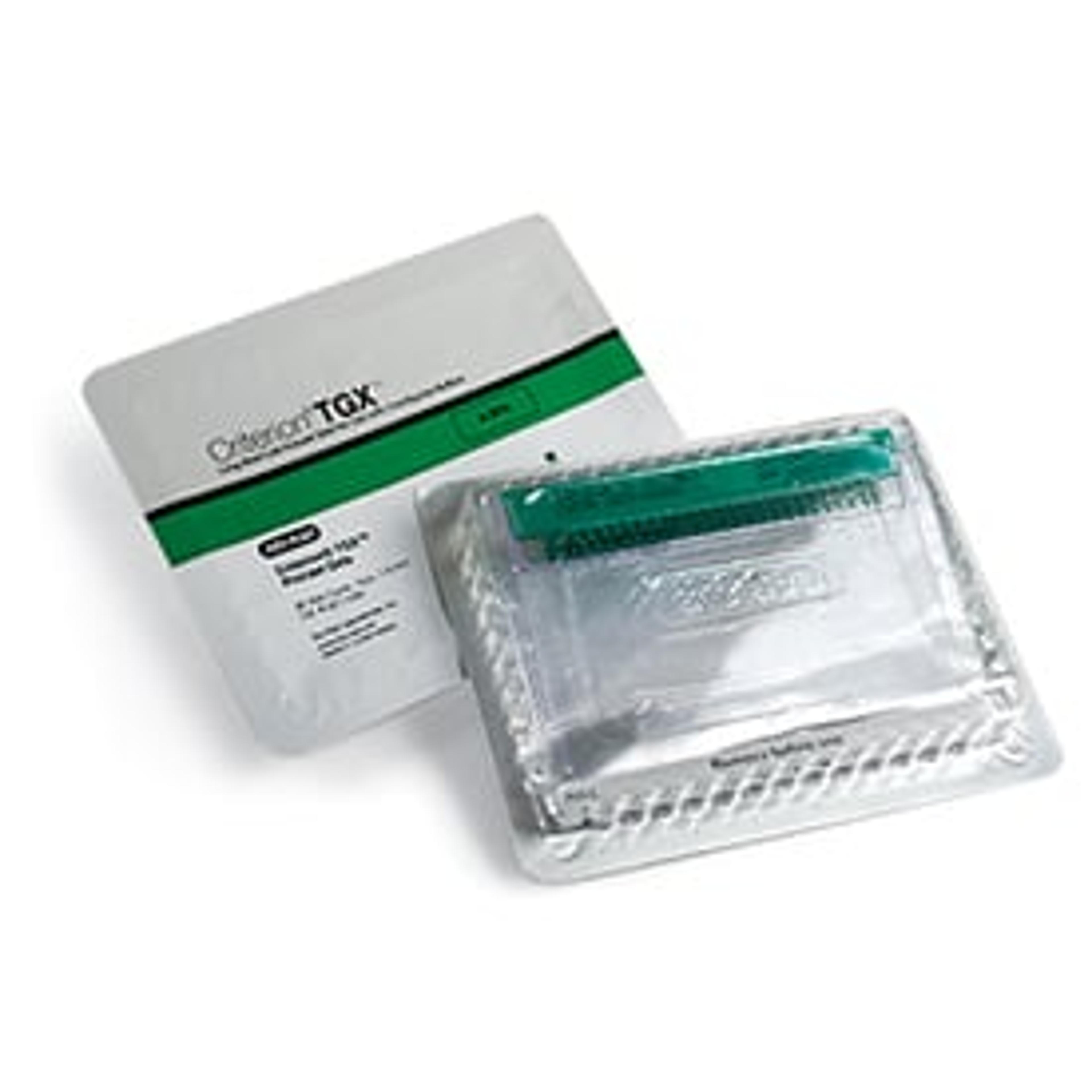
2. Separate proteins based on molecular weight (MW) by SDS-PAGE on TGX™ precast gels (the second dimension of 2-DE). The unique chemistry of Bio-Rad’s TGX gels enables fast electrophoresis times (less than 30 minutes) and highly efficient protein transfer.
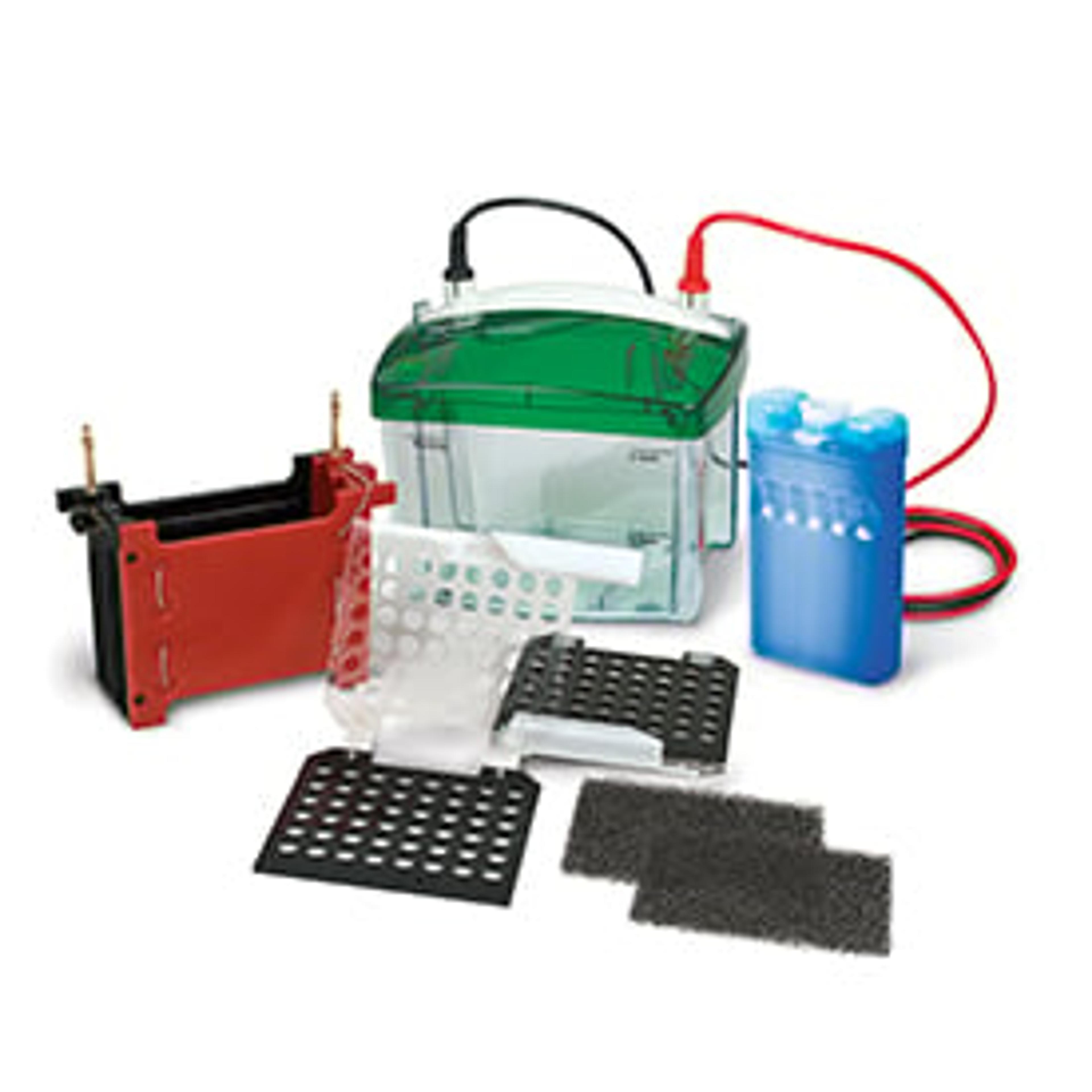
3. Process the gel with the Trans-Blot® Turbo™ system to transfer the 2-DE separated proteins to a nitrocellulose or PVDF membrane in less than 10 minutes. The unique combination of TGX precast gels and the Trans-Blot Turbo system enables highly efficient and reproducible transfer of proteins across a broad pI/MW range, which is essential for confidence in 2-DE/WB results.
4. Perform fluorescent protein staining with SYPRO Ruby (to confirm the efficiency of protein transfer) and western blotting using polyclonal anti-HCP antibodies on a nitrocellulose or PVDF membrane.
5. Image using the ChemiDoc™ MP imager and analyze with PDQuest™ software to determine the percentage of matching spots between those detected by membrane-bound protein staining and those visualized by immunodetection.
“Bio-Rad’s solution provides a more sensitive means to confirm the effective transfer of all proteins from the gel to the membrane, which is a crucial step in this study,” Ritter said. “If you see a low degree of immunoreaction on the blot, you won’t know if it is due to limited coverage of the antibodies or the absence of immobilized protein. Bio-Rad’s system assures that the proteins will be present on the blot.”


