Capture Protein Expression Changes Over Time in Living Cells
Learn how live-cell immunocytochemistry offers a much-needed temporal element to measuring protein expression in real time.
22 Aug 2018
Immunocytochemistry (ICC) as a basic cell imaging technique has been engrained into every cell biologist’s repertoire. Fluorescently labeled antibodies added onto fixed cells help visualize protein expression in the cells. While traditional ICC yields information on protein localization i.e. where the protein of interest is expressed, a big caveat remains – it is unable to capture protein expression changes over time, the ‘when’ of cellular dynamics.
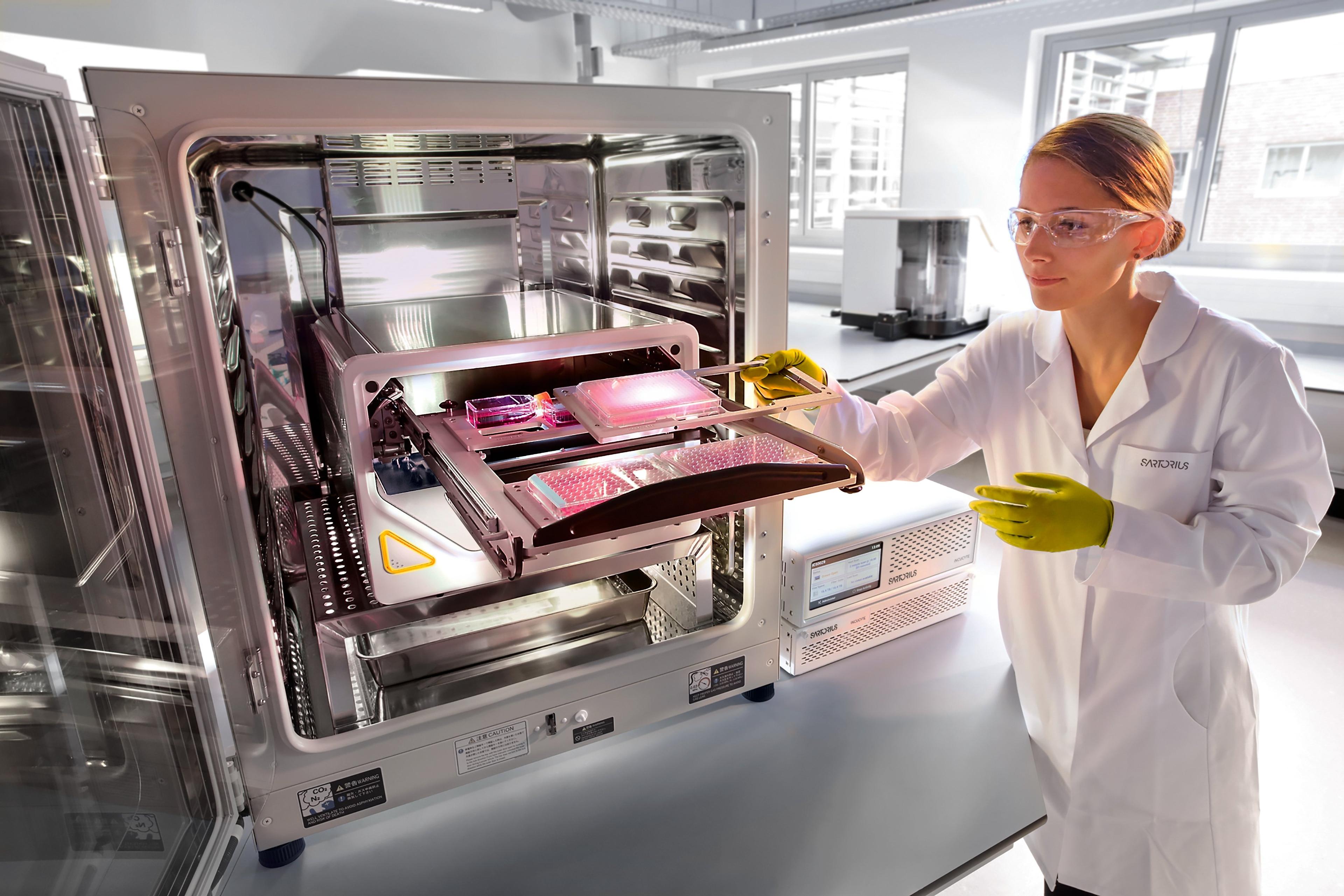
Along with the spatial resolution offered by conventional ICC, there is a need for temporal resolution that can reveal cellular states and cell-cell interactions, phenomena that cannot be visualized in fixed cells. Offering a way to capture cellular dynamics on live cells, while still keeping the benefits of ICC, Essen BioScience recently launched the IncuCyte® Live-Cell Immunocytochemistry.
Tim Dale, Director of Biology, Europe, at Essen BioScience speaks with SelectScience® to explain how live-cell ICC works, the importance of automated live-cell imaging and how it can improve your experiments.
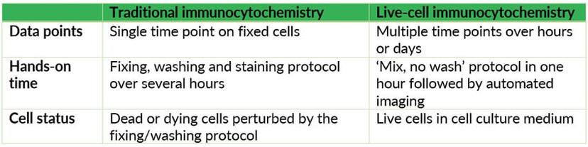
“ICC, in general, has been well established in most labs,” says Tim. “That being said, traditional ICC is limited when trying to study the changes in protein expression over time and really understand the dynamics of the process.” Live-cell ICC provides a solution to this unmet need, opening up possibilities for tracking protein expression in real time.
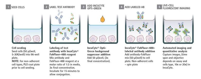
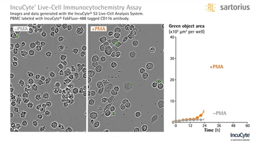
Live-cell ICC on the IncuCyte platform draws on the strengths of the instrument’s live-cell analysis capabilities, coupled with conventional fluorescently-labeled antibody binding. Using a simple mix, no-wash protocol, the antibody of your choice is first tagged with a fluorescently-labeled antigen-binding fragment. This is then added directly to your living cells, setting the stage for your live-cell ICC imaging. Adding in a background suppressor such as the IncuCyte Opti-Green helps obtain clean signals.
The removal of several wash steps and the ability to let experiments run while the IncuCyte automatically takes images over hours or days makes this a highly productive experimental set-up. “In addition to that, the automated imaging and analysis by the IncuCyte really enable you to do this in a 96-well plate,” says Tim. “The workflow is amenable to higher-throughput questions.”
“This has been designed with the flexibility of scientists in mind. So, you can take your choice of test antibody and then easily label it with the dye to identify the cells,” says Dr. Nicola Bevan, Principal Scientist at Essen BioScience.
“Live-cell ICC allows surface expression dynamics to be monitored. More importantly, these protein expression changes can be coupled to morphology and function by using this alongside many of IncuCyte’s functional applications,” says Tim. In a proof-of-concept experiment coupling protein dynamics to morphology and ultimately function, Nicola and her team measured the changes in key macrophage markers following two monocyte differentiation paradigms. They observed temporal increases in protein expression and critically marked differences in profiles depending on treatments. Furthermore, protein expression profiles could be related to the phagocytic potential of the macrophages, the key function of this cell type.
“You don’t have to fix and stain or do anything complicated with this experimental design. This lack of perturbation maintains the cellular environment which allows us to capture cell-cell interaction and quantify those interactions using live-cell ICC,” adds Tim. “This has the potential to reveal insights into the interplay between different cells within a micro-environment.”
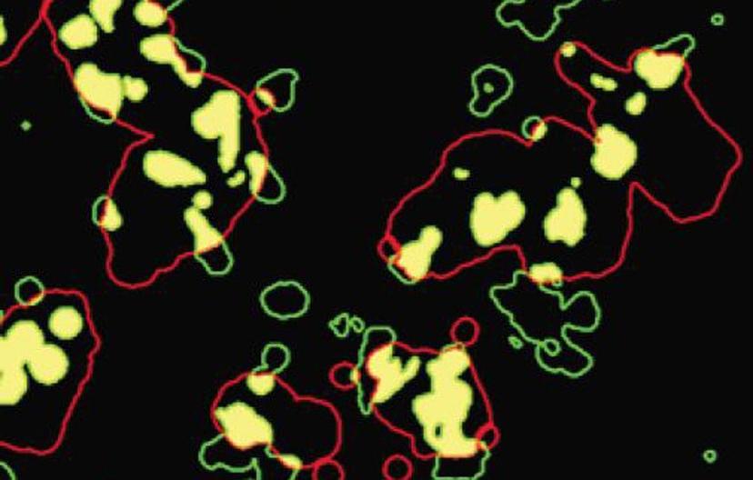
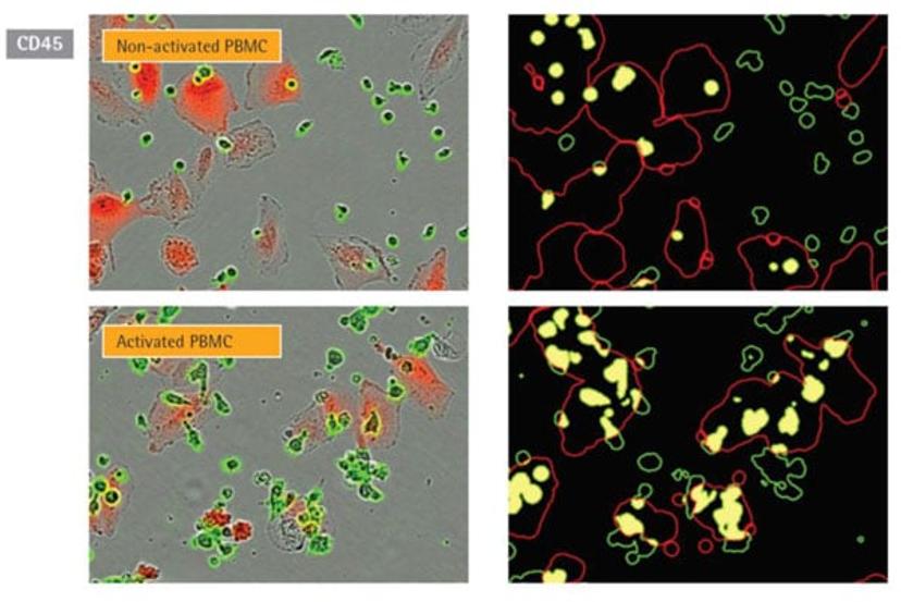
Nicola and the research team at Essen BioScience monitored cell-cell interactions between lung cancer cells and tumor cells employing live-cell ICC. RFP-expressing lung cancer cells were co-cultured with immune cells tagged with fluorescently labeled antibody, CD45. The IncuCyte analysis software created expanded image masks around the RFP signal. By quantifying the overlap of red (tumor cells) and green (immune cell) signals, it was possible to measure the interaction between the two cells.
“It has been exemplified using the live-cell ICC solution – being able to temporally monitor the cellular processes whether that’s through protein expression, cell health or function – really affords insights into cell physiology, pathophysiology and potential mechanisms of drug actions,” Tim explains. “Anything that modulates cell function can benefit from temporal analysis.”
Plus, the multiplexing potential of automated live-cell imaging yields data-rich experiments from even a single well. Nicola adds: “The IncuCyte has provided scientists with a workflow that makes it far easier to get more out of each well and each assay, because you don’t have to fit everything into your working day: the IncuCyte works even at night!”
“We take a very application-centric view towards the development of new products,” Tim says. “It’s better for a scientist to be able to focus on the biological questions than on technical solutions.”
As a highly reviewed product, that received a SelectScience Silver Seal of Quality this year, the IncuCyte has revolutionized the way cell culture experiments are designed and enjoys a collaborative customer relationship. “We get asked more routinely about the validity of reagents and its suitability for use in live cell analysis. It’s not always that common for general reagents to be suitable for live cell analysis,” Tim explains. “So, being able to provide additional information regarding the validity of our reagents is key. That’s why we endeavor to link validated reagents as a part of our integrated solutions.”
The next big step for live-cell imaging is tackling 3-dimensional translational models. “We are trying to develop applications to monitor 3D structures. We really want to aid the use of more relevant cell types whether they are iPSC-derived cells or patient-specific tissues,” says Tim.
Essen BioScience aims to offer simplistic features to study the complex cellular models of the future. “As 3D models get more complex, the cell types are likely to become more heterogeneous. So we are starting to think about how we can embrace that heterogeneity and track it over time. But we want to make the solutions simple for the end user: we will still provide all aspects of the workflow to add value to your science.”
Do you use the IncuCyte? We’d love to hear from you. Please leave a review here.