Collaboration between University of Manchester and Phasefocus™ Kicks Off with Livecyte Training
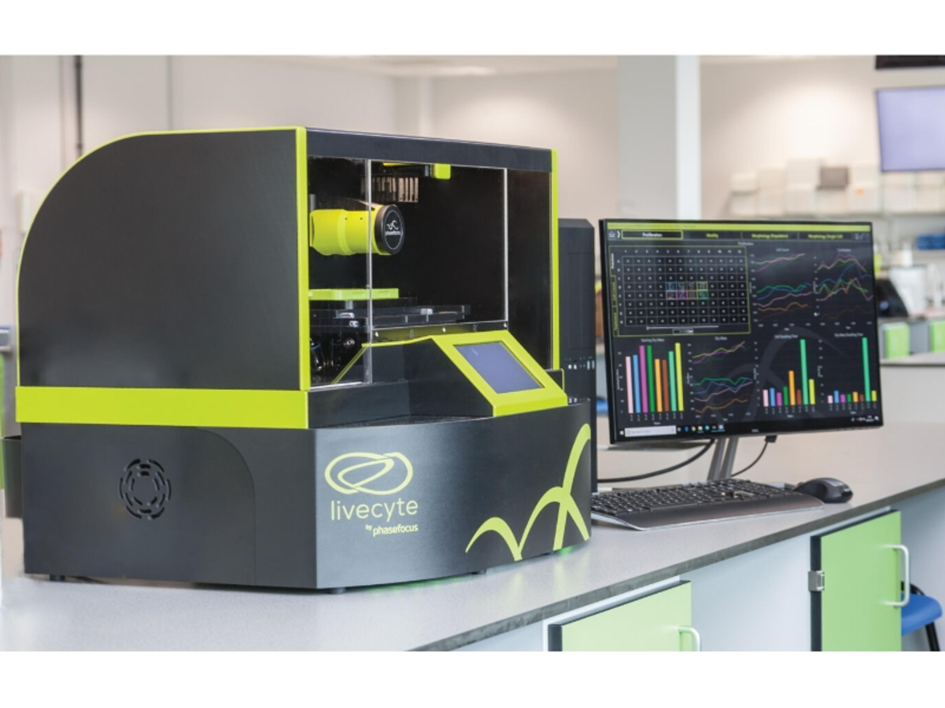
Following a successful funding application, staff at the University of Manchester, Wellcome Trust Centre for Cell Matrix Research, have become the proud owners of a Livecyte™ Cell Imaging & Analysis System
9 Nov 2017Livecyte is a unique quantitative label free cell analysis system which delivers continuous tracking and analysis of individual cells within large populations, and multiple wells, enabling users to gain a more realistic and comprehensive understanding of cell behavior.
The system, located at the University’s Bioimaging Core facility, has been specifically designed to create an optimal environment for long term live cell assays, minimizing phototoxicity and affording researchers the capabilities to compare normal and diseased cells in an increasingly natural environment.
The purchase of Livecyte heralds the start of a BBSRC CASE collaboration between the University of Manchester and Phasefocus, the Sheffield optical technology company who developed and manufacture the system.
The BBSCRC funded program aims to reinforce links between academia and industry by utilizing existing academic resources to support and develop innovative ideas from SMEs as a means of disseminating scientific knowledge to build best practice.
To see more lab informatics news, visit our Lab Informatics Community >>
Dr. Christoph Ballestrem, academic lead for the bioimaging facility commented “Livecyte is the perfect system for long term imaging and will allow us to gain a better insight into how cells sense and communicate with their environment and how this impacts tissue formation in both healthy and diseased states”. Determining the differences between healthy and diseased cells is at the heart of Dr. Pat Caswell’s research interests. Together with Dr. Ballestrem and Prof Tim Cootes, Pat is part of the interdisciplinary supervisory team and believes that the new system can help identifying behavioral signatures between cells.
In return, Phasefocus hopes to benefit from the expertise of the University’s Imaging Sciences group, headed by Prof Tim Cootes, in the development of algorithms to analyze the images. Machine learning techniques will be used to automatically detect cells and to measure their behavior.
PhD student, Tristan Henser-Brownhill, will spend 3 months in Sheffield with Phasefocus as part of the project. Outlining his incentives for participating in the scheme, he stated “This is one of the first microscopes that can accurately detect and quantify dry mass. The use of quantitative phase imaging (QPI) can detect small changes which we can correlate with fluorescence markers and the computational power to apply this technology for practical applications has only recently become available. Phasefocus is the first company to make that happen”.
Dr. Egor Zindy also added “Phasefocus have been very receptive and responsive to suggestions from end-users and researchers are pleased to know that the product is being designed and developed to meet their needs - and very quickly.”
Dr. Rakesh Suman, from Phasefocus responded “We have an opportunity to refine the direction of our development work, based on feedback from Livecyte customers and collaborators. This is an exciting time for Phasefocus as we can learn a lot in a very short time with a project like this”