Doubling the resolution of structured illumination microscopy
New image reconstruction algorithm enhances super-resolution microscope ZEISS Elyra 7
29 Apr 2021
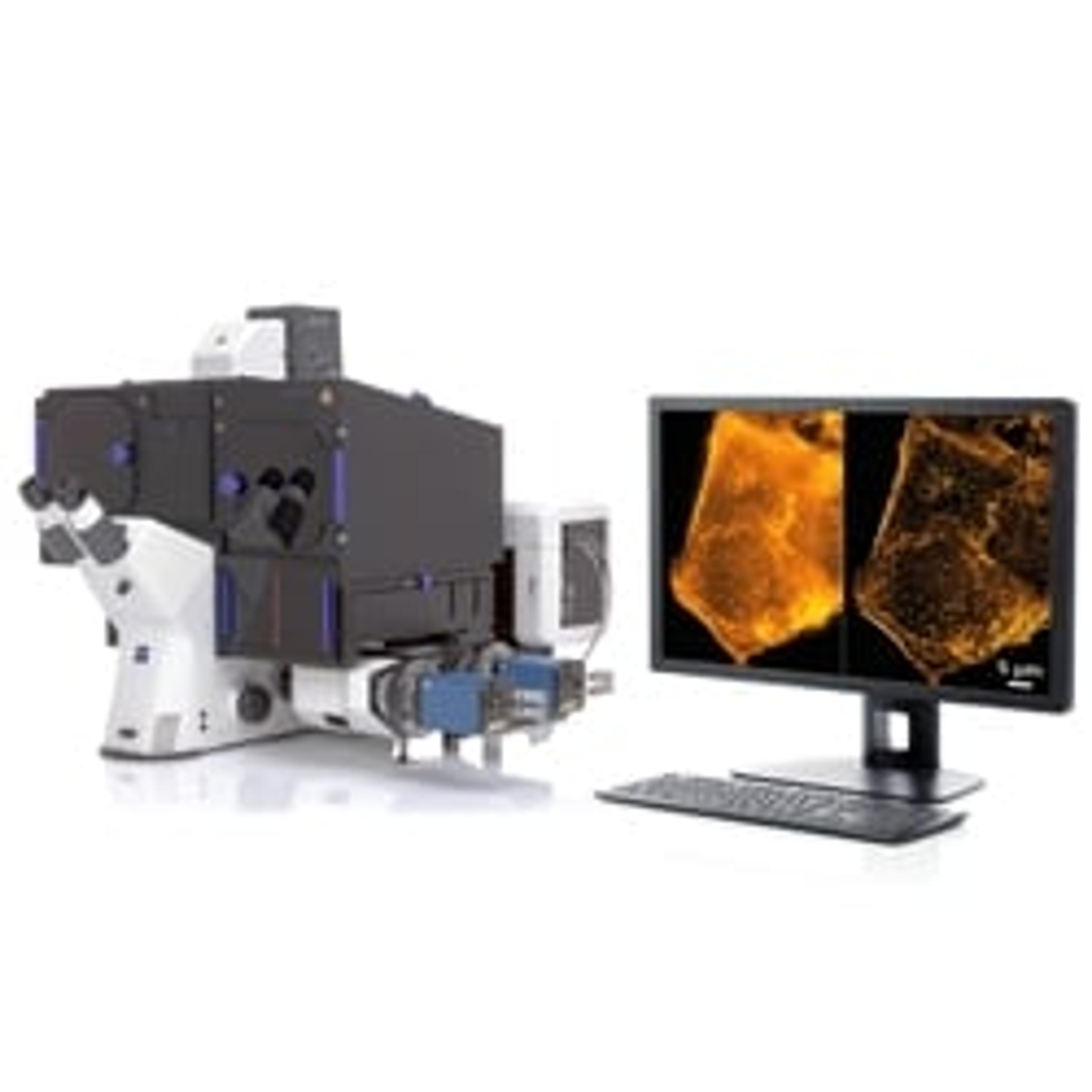
ZEISS has introduced SIM², a groundbreaking image reconstruction algorithm that increases the resolution and sectioning quality of structured illumination microscopy (SIM) data. With SIM² on the microscope system ZEISS Elyra 7, life science researchers can now double the conventional SIM resolution and discriminate the finest subcellular structures of living and fixed samples, even those no more than 60 nm apart.
SIM is an established grid-based illumination technique that allows imaging at resolutions beyond the diffraction limit of optical microscopy. With the introduction of ZEISS Lattice SIM two years ago, ZEISS went a step further and combined the resolution advantages of SIM with a dramatic increase in imaging speed and detection sensitivity, making the super-resolution microscope ZEISS Elyra 7 a preferred choice for live-cell imaging. With SIM², the consistent efforts to further develop the ZEISS super-resolution technology are now taking another step further, allowing researchers to exceed previous limits of resolution, imaging speed, and gentleness.
New algorithm enhances resolution, sectioning, and robustness
The new reconstruction algorithm SIM² is superior to conventional image reconstruction methods in terms of resolution, sectioning, and robustness – all without the need for tailor-made staining protocols or expert knowledge of complex microscopy techniques. SIM² not just resolves structures down to 60 nm but allows super-resolution and high-dynamic imaging at the same time – indispensable necessities to observe fast biological processes in living cells or organisms.
Living biological samples at resolutions well below 100 nm
With SIM², researchers can now, for the first time, view details of living biological samples at resolutions well below 100 nm – with speeds of up to 255 frames per second. The ease of access to this high spatiotemporal resolution will enable new discoveries of subcellular functional principles and contribute to a better understanding of the distribution and architecture of organelles. Researchers in developmental biology, neuroscience, plant sciences, and neighboring disciplines will gain more insights into their model organisms and specimens by revealing fast cellular processes, resolving structures in 3D with high penetration depth, and investigating structural changes at the molecular level.
Customers who have been participating in a beta test program immediately recognized the potential of ZEISS Elyra 7 with Lattice SIM² for their research and expressed enthusiasm in view of the new possibilities. Peter O’Toole, Head of Imaging and Cytometry at the University of York: “I remember seeing the first results. I could only laugh as I was simply amazed. My next reaction was to email some of the key users who could immediately benefit. From the tissue neurobiologists, to cell and molecular immunologists, to those working with yeast and those working with bacteria, all of them are already gaining from SIM².”
With the introduction of SIM², ZEISS is taking the next step to continuously develop ZEISS Elyra 7 into the major platform for live cell-compatible super-resolution microscopy. This underscores the ZEISS drive to make advanced imaging technology easily accessible to the scientific community.
Want the latest science news straight to your inbox? Become a SelectScience member for free today>>