Establishing Phantoms to Improve MRI Signal and Quality
SelectScience® spoke to Dr Michael Boss, NIST, about his work to improve brain imaging
19 Feb 2016

SelectScience® spoke to Dr Michael Boss, NIST, about his work to improve brain imaging

National Institute of Standards and Technology (NIST) NIST is a measurement standards laboratory, promoting U.S. innovation by advancing science, standards, and technology.
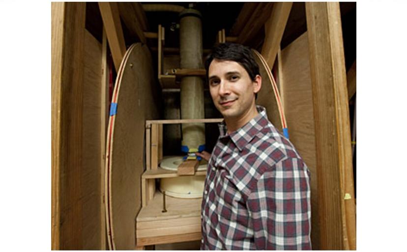
Nuclear magnetic resonance (NMR), is a physical phenomenon that can reveal detailed information on the chemical structure of unknown compounds, accurately measure purity, and, most commonly, be utilized in magnetic resonance imaging (MRI) for medical diagnosis. However, MRI information can be distorted or littered with artifacts, which lessen the certainty of a diagnosis. SelectScience spoke to Dr Michael Boss, of the Magnetic Imaging Group at NIST and specializing in brain imaging, who is working on reducing these interferences.
“Brain imaging provides a challenge”, Dr Boss explains; “neural tissue is very complex, as is the geometry of the head”. He is working towards the development of quantitative reference standards for MRI, and the establishment of a ground truth. “Ground truth is the true value of a physical parameter”, and without it, there is “no guarantee that the number measured is the true value”, he explained. Using the “gold standard” techniques of NMR, the accurate measurement of the ground truth will allow accurate measurements of all kind of parameters.
Eliminating distortions with phantoms
The standards Dr Boss is developing are known as “phantoms”. They are reference objects with known properties and can establish the “geometric distortion of an MR image, the signal-to-noise or contrast-to-noise ratio, or the spatial resolution”. The goal of his group is to “establish quality control protocols that enable reproducible, quantitative MRI. For example, if you were looking to generate a spatial map of the diffusion coefficient in a cancerous tumor, having a phantom with known diffusion properties is paramount to obtaining an accurate map in vivo.”
Magnetic resonance provides such an abundance of information that there exist almost limitless possibilities for harmonization with other spectroscopic and imaging techniques. Combining NMR/MRI with optical spectroscopic techniques could provide “a wealth of information on living subjects – using complementary but physically different techniques” allows for confident cross-validation. NIST is interested in combining MRI with positron emission tomography (PET) as a standard for multi-modality imaging. They are also planning on using microCT (another imaging technique utilizing x-rays) as an independent means of validating some of their phantom structures.
The future of MRI
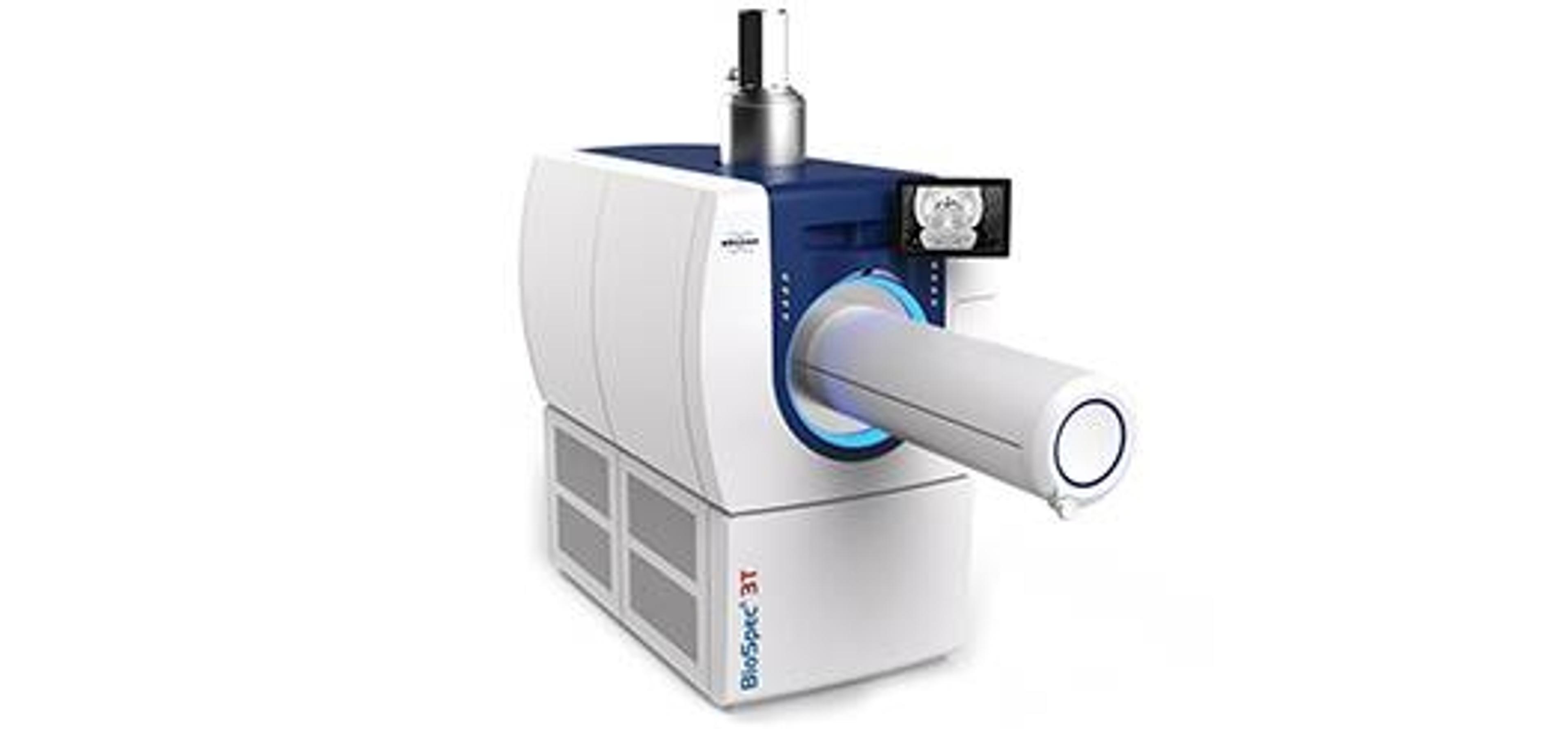
“There are currently strongly efforts to push MRI to strengths of 7 tesla and beyond”, Dr Boss revealed. At present, MRI is clinically performed at 1.5 and 3 tesla, and although going to higher field strengths increases the signal, “it also introduces new problems in terms of image quality”. However, if these problems can be overcome, “these new MRI scanners could have higher spatial resolution, provide new means of generating contrast in an MR image, enable better MR spectroscopy, and lead to improved patient care through better diagnostics.”
Learn more about NMR and spectroscopy on our dedicated page, or read about how NMR can be utilized in other applications.

