Editorial ArticleLife Sciences
How to achieve high-quality imaging of neurological tissues
Learn how one scientist is harnessing the latest imaging technology to advance his research and generate high-resolution images of neural tissue
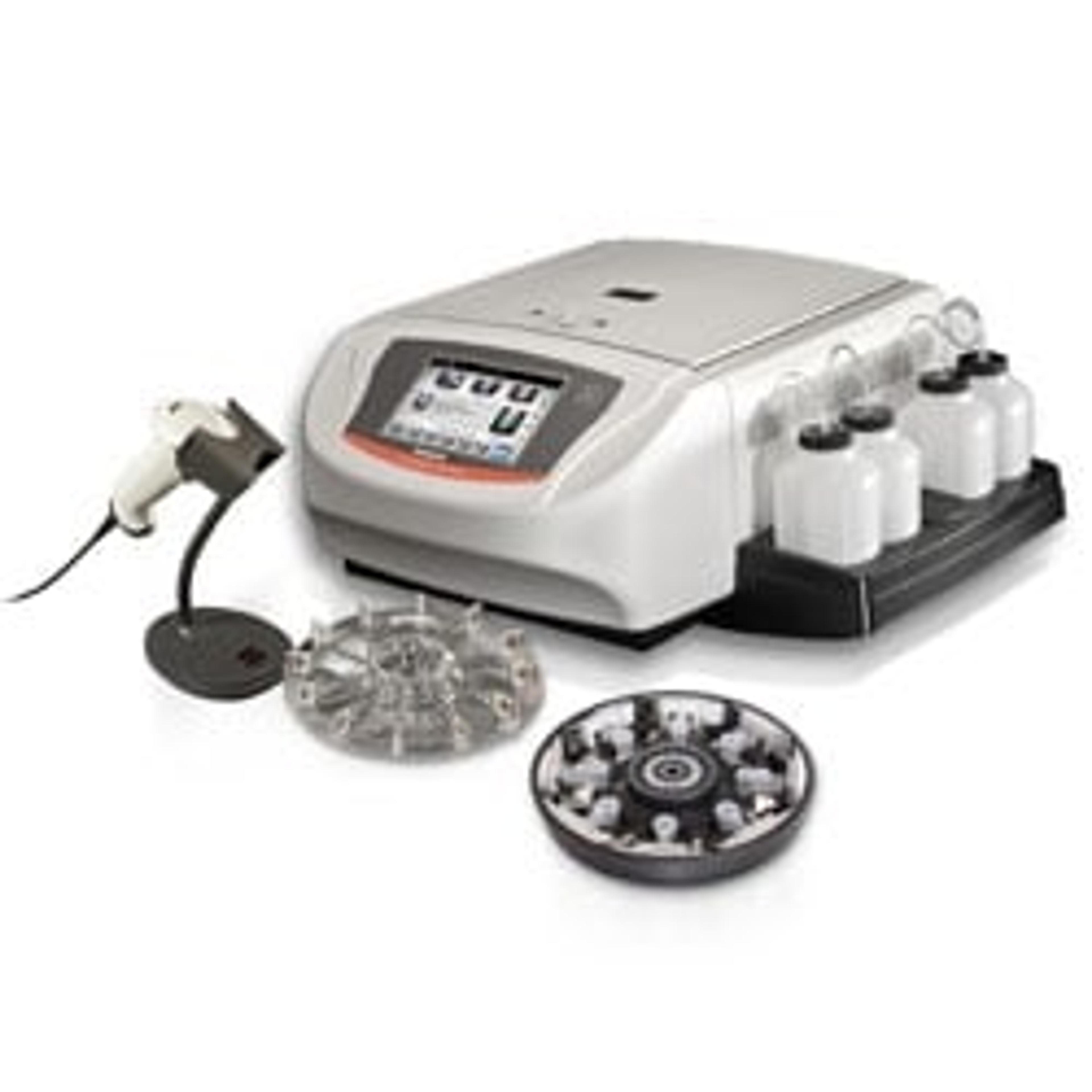
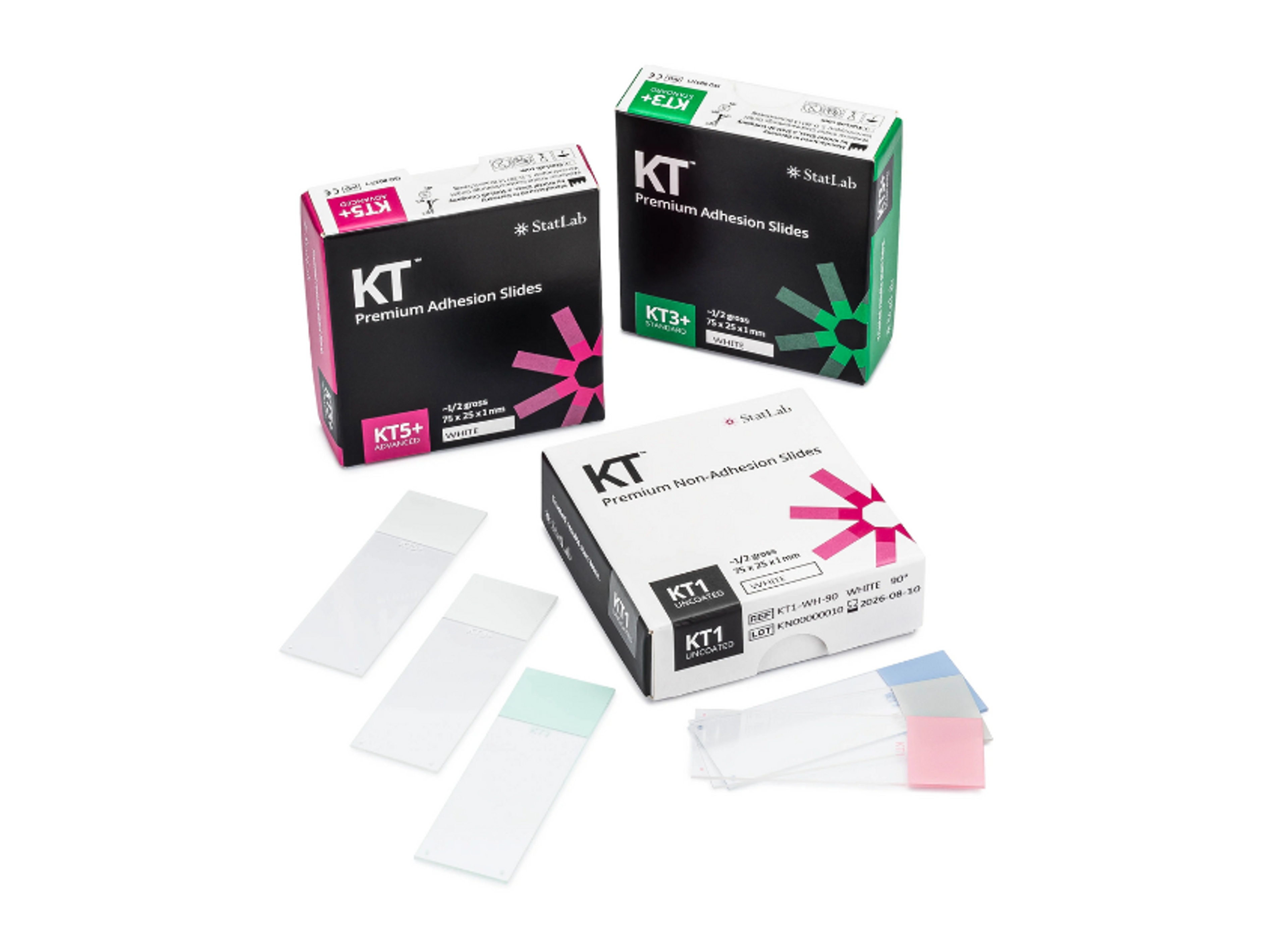
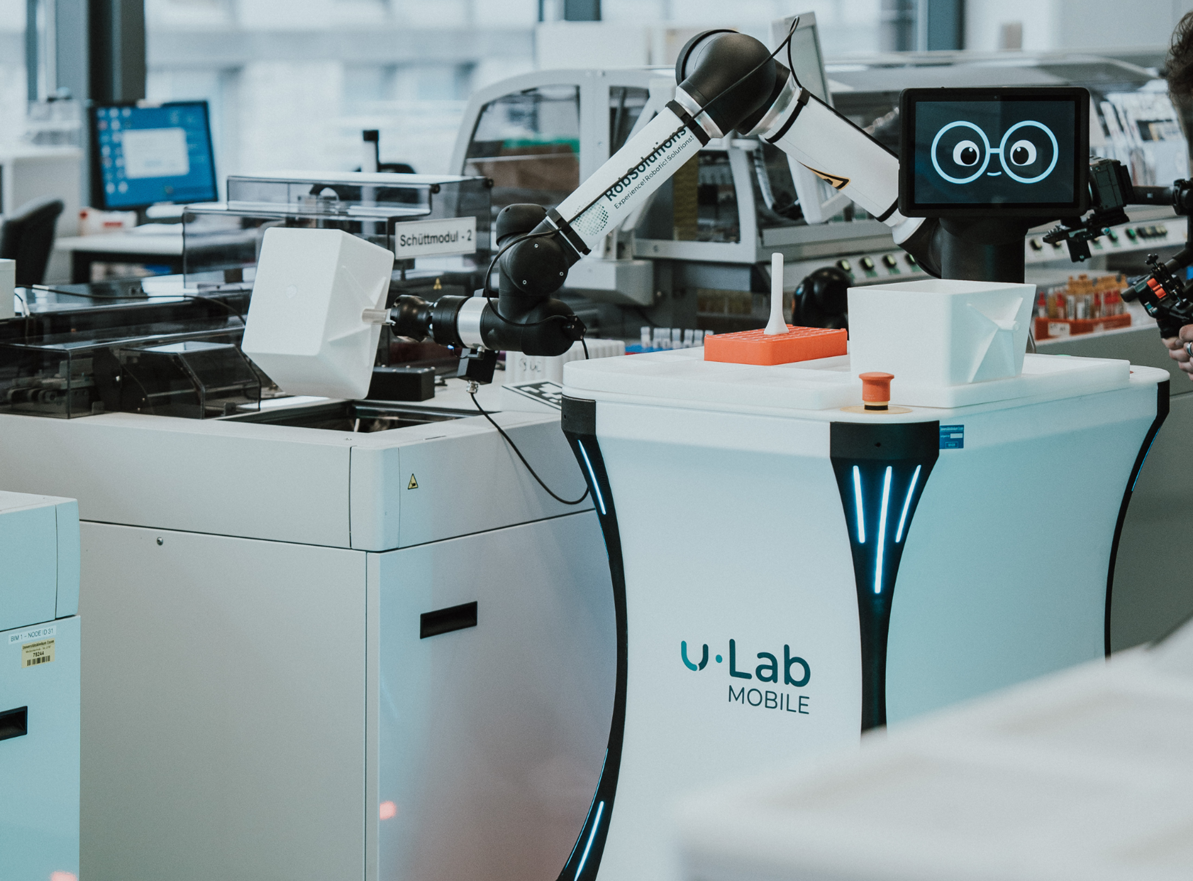
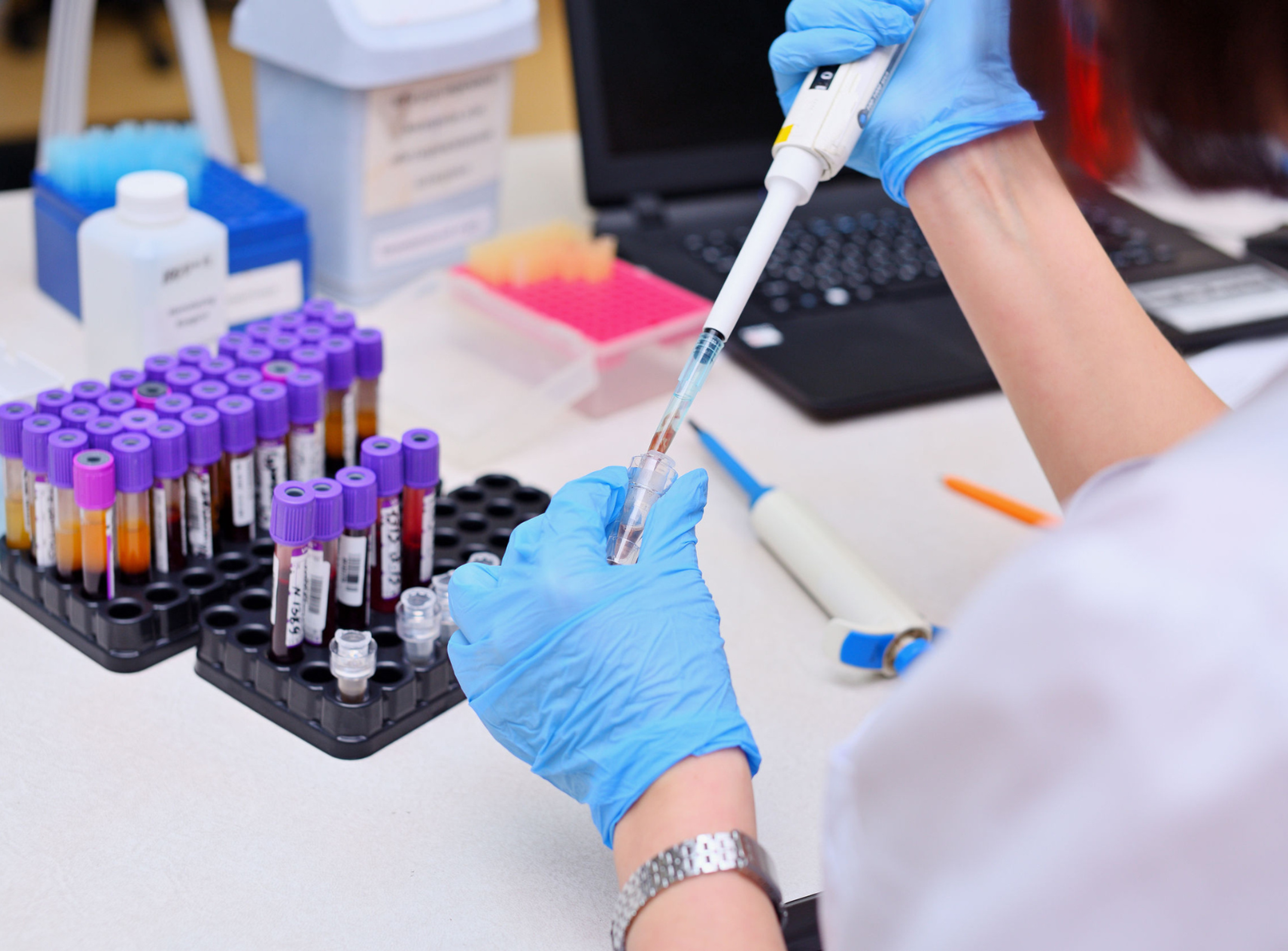
Cellular Pathology deals with the microscopic analysis of tissue samples and cells. Sample preparation and processing includes fixation, staining, sectioning and slide mounting, using equipment such microtomes and cryostats. In choosing immunohistochemistry and immunocytochemistry kits, consider chromogens, staining method, antibodies, microscopes and imaging.
Learn how one scientist is harnessing the latest imaging technology to advance his research and generate high-resolution images of neural tissue
Great piece of equipment, highly recommended!
The ability to designate multiple points for observation simultaneously is very convenient