Game-changing microscope redefines single-cell science
Find out how to unlock cellular mysteries with Yokogawa’s Single Cellome™ System SS2000
16 Jan 2025
Studying individual cells can reveal insights that bulk analyses might hide, from understanding cellular responses to mapping molecular details within distinct cell populations. Recently, Yokogawa’s Single Cellome™ System SS2000 has emerged as an innovative tool in single-cell analysis, allowing researchers to study cellular morphology, spatial patterns, and molecular interactions simultaneously, all within the same live cells.
SelectScience® spoke with Dr. Johanna von Gerichten and Dr. Jake Penny, research fellows at SEISMIC, a BBSRC-funded facility dedicated to single-cell and subcellular omics, who shared their experiences and insights on this cutting-edge instrument. Together, their work with the SS2000 is helping scientists better understand cellular heterogeneity and unlock new layers of biological complexity.

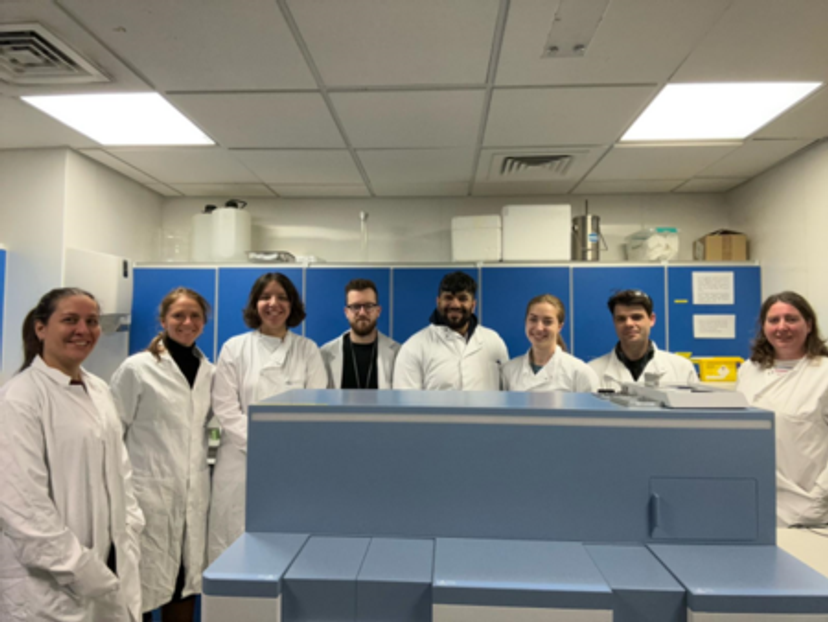
The SEISMIC team at University of Surrey
How to bridge imaging and molecular analysis
The Yokogawa SS2000 is a dual microlens spinning disk confocal microscope designed to seamlessly integrate microscopy with advanced analytical techniques, such as mass spectrometry and sequencing. This technology enables researchers to study living cells over time in a controlled, incubated environment.
For scientists like von Gerichten and Penny, this has opened up entirely new approaches to cellular analysis. "The most exciting part of this instrument is that you look at a living cell culture and can decide, ad hoc, ‘I want this cell now that behaves like this at this time point,’” von Gerichten explains.
This flexibility is a breakthrough for researchers exploring cellular behaviour. Single cell sampling is still a novel method, however, many methods of single cell isolation requiring removing the cells from its natural state leading to cell stress that can potentially skew results. The SS2000, however, keeps cells alive and in their natural state, allowing scientists to capture time-based changes in cellular morphology and lipid composition with minimal disruption. With this approach, scientists could observe living cells over extended periods and capture intricate cellular events as they unfold, such as signal transduction, migration, and membrane dynamics.
Unmatched versatility for high-content imaging and single-cell sampling
A standout feature of the SS2000 is its ability to provide both spatial and morphological data for each cell sampled, a dual capability that sets it apart from traditional cell sorters. “It’s not just single-cell picking and single-cell analysis,” says Penny. “We’re able to also gain spatial and temporal data, which lets us look at a whole range of different biological processes.”
This system integrates Yokogawa’s renowned CSU W-1 Nipkow spinning disk confocal technology, which minimizes phototoxicity and facilitates time-lapse imaging, allowing researchers to monitor cells over time without causing damage. The SS2000 also includes a high-precision stage incubator, capable of controlling the environmental conditions of sampled cells. Such features are crucial for researchers studying dynamic cellular events, enabling them to draw a more holistic view of each cell and understand cellular processes as they occur naturally.
Adding to its versatility, the SS2000 comes with various sampling tip sizes (10µm, 8µm, 5µm, and 3µm), allowing researchers to work with a wide range of cell types. Furthermore, it supports a broad range of objectives, from 4x to 40x, which are essential for achieving high-resolution images suited to diverse cellular studies. These customizable parameters make the SS2000 adaptable for different research needs, from proteomics and lipidomics to genomic analyses.
A flexible and user-friendly tool for collaborative research

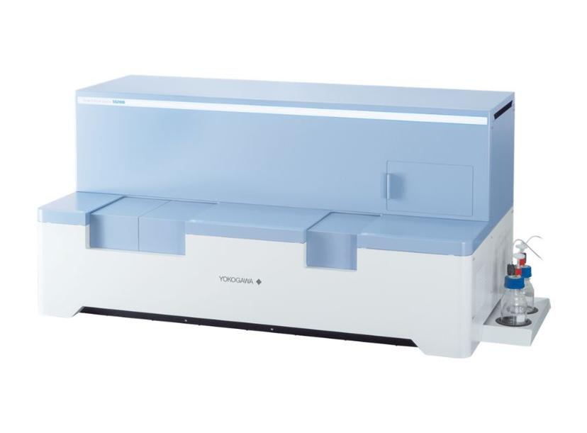
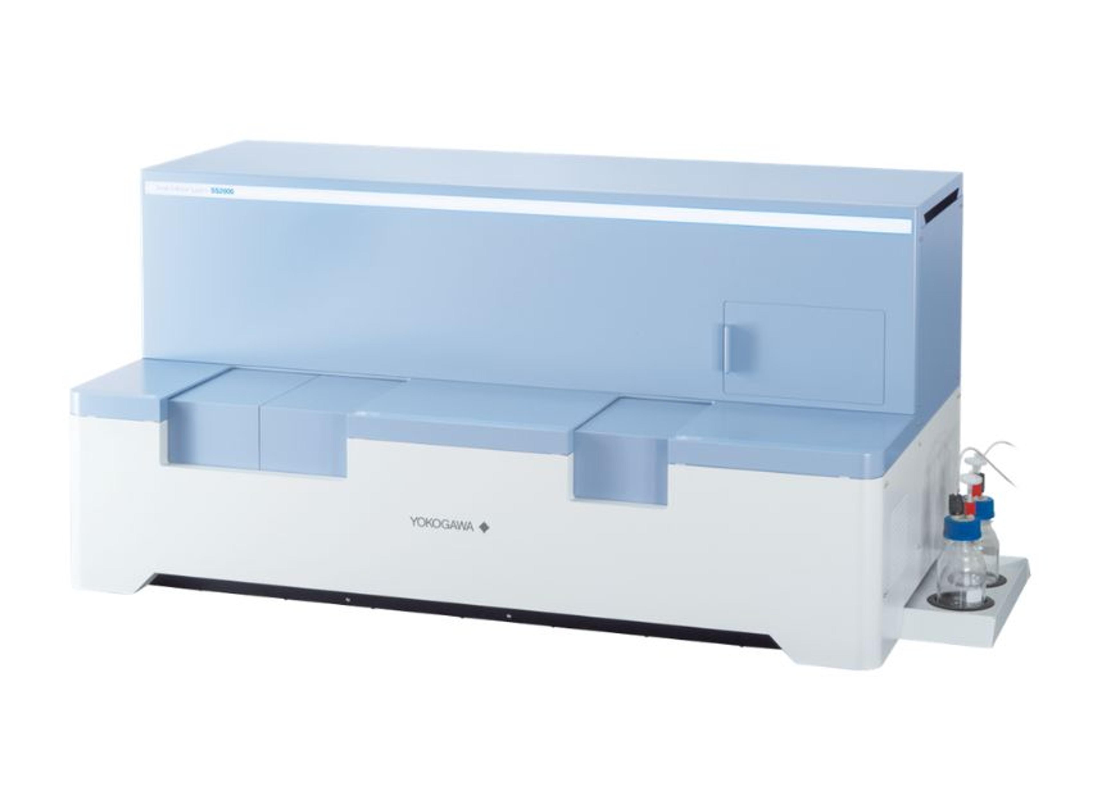
The Yokogawa Single Cellome™ System SS2000 integrates Yokogawa's renowned spinning disk confocal microscopy technology with a sophisticated sampling mechanism
Another significant advantage of the SS2000 is its ease of use, which has encouraged researchers from various fields to explore single-cell studies. Penny notes that it doesn’t require extensive technical training, making it accessible for collaborative projects: “it doesn’t need a specialized skill set; just the right amount of caffeine for steady hands!”
The SS2000 has become a cornerstone in SEISMIC’s collaborative work, where biologists, biochemists, and analytical chemists alike can contribute to and benefit from its capabilities. This accessibility is further enhanced by Yokogawa’s commitment to supporting users through regular visits and responsive feedback loops. “We are in constant communication with them,” von Gerichten notes, emphasizing the productive partnership that has helped them optimize the SS2000 for a wide range of applications.
This openness has been particularly useful for unexpected projects. For example, Penny recalls collaborations in virus and bacterial research, areas that neither SEISMIC nor Yokogawa initially anticipated as high-usage cases. Through collaborations like these, the SS2000 has demonstrated its value as a versatile instrument capable of addressing diverse research questions.
Improved understanding of mechanisms in health and disease
The SS2000 is contributing to significant advancements in health and disease research, particularly in areas that benefit from understanding cellular heterogeneity. By capturing both imaging and analytical data from individual cells, researchers are able to explore connections that were previously beyond reach.
In cancer research, for example, single-cell analysis has revealed that certain resistant cancer cell subpopulations can evade treatment, leading to recurrence. According to von Gerichten, capillary single cell sampling has been pivotal in investigating drug uptake in cancer cell and has sparked interest for single cell lipidomics and the SS2000 further streamlines the sampling workflow. “This research started off with drug uptake studies on single cells. Then we realized we could also see lipids,” she says, describing the pathway that has led to their current work with the SS2000.
Another promising application area is immune response and infection research. SEISMIC has partnered with collaborators interested in how cells react to viral infections, an area where the SS2000’s precise sampling and imaging capabilities have been invaluable. Using the SS2000, researchers can infect cells with specific viruses, then track cellular responses, such as the formation of stress granules—protein clusters that cells form under stress. As Penny explains, “These are quite unstable structures, and if you try to isolate them through traditional methods, they just dissolve.” With the SS2000’s capillary-based sampling, however, researchers can isolate these granules within infected cells and analyze them in detail, gaining insights into the cellular defense mechanisms that are critical to understanding viral infections.
The future of single-cell lipidomics and beyond
The SS2000’s contributions to cellular analysis are part of a larger shift in single-cell research. With the ability to track changes in individual cells and understand how cellular heterogeneity affects larger biological processes, scientists are reshaping their approach to lipidomics, immune response, and physiological studies. “Single-cell lipidomics is so new,” says von Gerichten. “It’s literally just a couple of years old.”
The field is rapidly evolving as scientists develop new methodologies for handling the vast data generated at the single-cell level. This is particularly relevant for studies where cellular-level differences can profoundly impact overall health.
For example, this field could soon explore the relationships between rare cell types and disease. “Researchers are interested in rare cell types within cardiovascular tissue and how these cells might help regulate surrounding cells,” Penny explains. This type of research could lead to unexpected findings, illuminating the role of rare cells in mediating tissue health or revealing potential therapeutic targets.
The versatility and precision of the SS2000 have also paved the way for lipidomics studies in aging and immune response. SEISMIC researchers are now investigating how cellular communication and bystander effects influence processes like inflammation, aging, and stress response. By using the SS2000 to track spatial and morphological changes in single cells over time, they are uncovering new patterns in cellular interaction and organization, which could have implications for understanding allergies, immune aging, and gut health.
In many ways, the work being done with the SS2000 is still laying the groundwork for future breakthroughs. As Penny notes, the team at SEISMIC is developing foundational methods that will allow them to address increasingly complex biological questions. “Once we’ve worked out all the nitty-gritty details, we can really start building up and asking exciting biological questions,” he says. With future applications ranging from disease model studies to drug development, it’s clear that this instrument has the potential to shape the landscape of single-cell research for many years to come.

