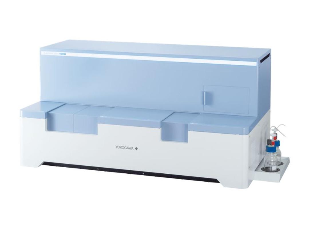
Single Cellome™ System SS2000
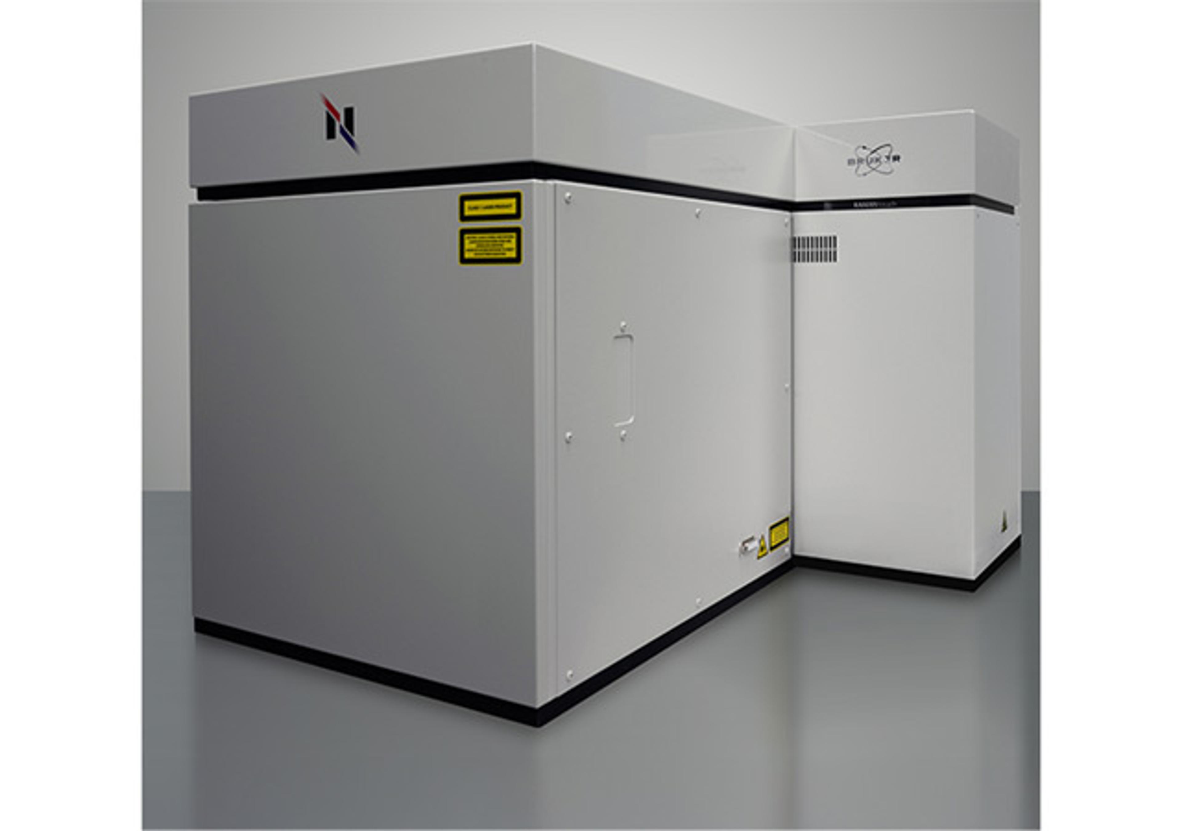
The Single Cellome™ System SS2000 is a dual microlens spinning disk confocal system for live-cell high-content imaging which can sample adherent single cells. Combining imaging with sampling ensures the preservation of spatial, morphological, and temporal information of samples for flexible downstream mass spectrometry and sequencing applications.

The supplier does not provide quotations for this product through SelectScience. You can search for similar products in our Product Directory.
At its core, the Yokogawa Single Cellome™ System SS2000 integrates Yokogawa's renowned spinning disk confocal microscopy technology with a sophisticated sampling mechanism. The SS2000 is a dual microlens spinning disk confocal microscope specifically designed to bridge the gap between imaging/microscopy and analytical techniques, such as mass spectrometry and sequencing, at the single-cell level. Researchers can observe and analyze dynamic cellular events, such as signal transduction, cellular migration, or membrane dynamics in adherent cells growing in an incubated environment. Once events of interest occur or target cells are located, the SS2000 can sample and collect them in vessels appropriate for analysis, such as PCR tubes and plates, all the while ensuring the preservation of spatial, morphological, and temporal information of the collected sample.
The SS2000 samples cellular material using micro-diameter capillaries of distinct sizes to accommodate for different cell sizes. The captured samples can be used in many different downstream analyses such as lipidomics, metabolomics, proteomics, transcriptomics, and genomics. The single-cell sampling capability is crucial for studies in which the analysis of individual cells and their heterogeneity can yield valuable insights, such as cancer cell analyses, studies of rare cell types, or personalized medicine. The SS2000 enables researchers to collect and image multiple cells from the same culture, and does not require cell suspension, which could induce cellular stress. This innovative system offers unparalleled capabilities for researchers seeking to correlate cellular behaviors over time and/or cell-cell interactions with intricate structural details.
Key Features and Benefits:
- Multiple sample tip sizes (10µm, 8µm, 5µm, 3µm) for flexibility in sample type selection
- Customizable sampling parameters for variable cell types
- Flexible collection sample holder options for various downstream sample analyses
- Environmental control of the sample collection chamber ensuring sample preservation
- High content imaging at your fingertips
- Collect high resolution images to facilitate sophisticated cellular image analysis
- Lasers supply coherent excitation light for shorter exposure times
- Supports a wide range (4x to 40x) of high-quality objectives, including phase and long working distance
- Up to 6 objectives can be installed simultaneously
- Environmental control of sampled cells with high precision stage incubator including hypoxia experiments
- No need to sacrifice image quality for speed with integrated CSU W-1 Nipkow spinning disk confocal technology
- Large field of view and tiling capability allows for specific choice of sampled cells
- Low phototoxicity of confocal images makes timelapse analysis possible
- Enclosed system removes the need for a dark/special room design while maintaining a centralized lab environment
Applications:
- High content imaging
- Single cell sampling for downstream single-cell omics analyses
- Lipidomics
- Metabolomics
- Proteomics
- Transcriptomics
- Genomics
- Link morphological, temporal and spatial information to data obtained from downstream analyses
- Montage/tiling of samples larger than camera FOV
- Capture fast biological events
- 3D images and analysis of spheroids, organoids and other complex structures
- Time lapse imaging
Not available in the following countries: North Korea, Iran, Iraq, Libya, Cuba, Syria, Sudan, South Sudan, Afghanistan, Democratic Republic of Congo, Central African Republic, Somalia, Lebanon, India, Pakistan, Russian Federation, Republic of Belarus, Ukraine.

