Glioma Stem Cells Enable the Production of 3D Human “Mini Brain” to Improve Glioblastoma Treatment
Discover how 3D models of glioblastoma using patient-derived stem cells could enable more effective treatment
24 Jul 2018
SS: Tell us a little about your background and your role at Weill Cornell Medicine (WCM)
AL: I have over 13 years of scientific training in a combination of government institutes and multiple premier academic medical centers including the National Institutes of Health, Vanderbilt University Medical Center and, most recently, New York-Presbyterian Hospital/Weill Cornell Medicine (WCM). I currently serve as the Director of the Starr Foundation Cerebral Organoid Translational Core here at WCM. In addition to establishing a “mini-brain” ex vivo model of glioblastoma (GBM) for our core, I manage the derivation of glioma stem cells (GSCs) from patient tumor tissue and oversee the compliance and key operations of our core facility.
SS: Why is there a need for organoids? What benefits do organoids offer over regular cell culture or even animal models?
AL: Current preclinical models of GBM are significantly limited by the lack of a normal human microenvironment. For example, animal models of GBM are restricted by the unknown interspecies differences between human GSCs and murine brain cells. Coupled with variably long tumor latencies and the inability to genetically manipulate and image tumor progression in real time, mouse models present a multitude of obstacles when trying to accurately reproduce the biology of human GBM. Tumor organoids overcome some of the logistical constraints of animal models, but they fail to address the important issue of cell-cell interactions between tumor cells and a normal host microenvironment.
SS: Tell us about your glioma cerebral organoid model
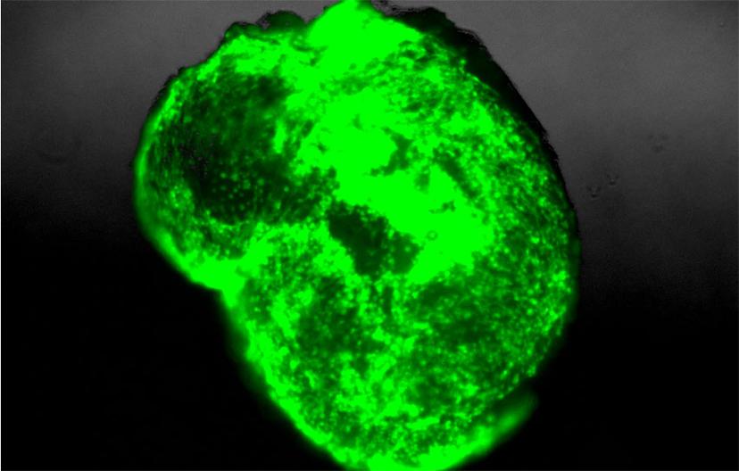
AL: To address the limitations of current preclinical GBM models, we have retro-engineered patient-specific GBMs using patient-derived glioma stem cells and human embryonic stem cell (hESC)-derived cerebral organoids. Our cerebral organoid glioma (GLICO) model demonstrates a clear infiltration of the human cerebral organoid by the patient-derived GSCs. Following the invasion into the organoid, the GSCs then proliferate and migrate throughout the host tissue, ultimately forming tumors that closely phenocopy patient glioblastomas.

SS: Please briefly describe your project using the glioma cerebral organoid model and share your findings
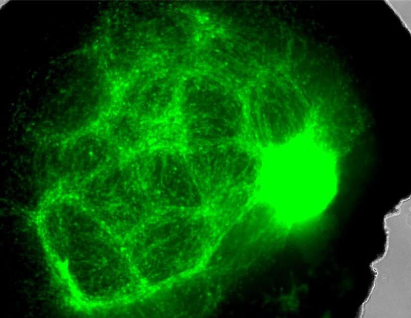
AL: Our GLICO model addresses a number of the limitations of current preclinical GBM models, as it allows us to study patient-specific GBMs within a human microenvironment similar to that of a normal human brain. Additionally, our cerebral organoids cultured and differentiated in Corning Matrigel matrix, allows us to recapitulate the same biological behavior and histopathological features of patient GBMs. Our findings also show that GLICO tumors are supported by a network of interconnecting microtubes that facilitate the invasion and proliferation of the glioma stem cells within the organoid.
SS: How important is it to use the right labware, surfaces and reagents to obtain an organoid culture?
AL: Selecting the appropriate labware is vital to generating a successful batch of cerebral organoids. As with any biomedical research involving human samples, it is critical that our data is consistent and reproducible each and every time. Failure to use the correct culture vessels, matrix or reagents can drastically alter the morphological structure and viability of the organoids. Corning® Matrigel® matrix is an integral component of every stage of organoid development in our lab. From culturing the hESCs and generating neuroectoderm/neural rosettes, to ultimately providing a three-dimensional matrix that promotes the growth of a fully differentiated cerebral organoid, Matrigel matrix is an indispensable component of our organoid research.
SS: What’s the future of organoid research? What projects do you have in the pipeline?
AL: The future of organoid research is incredibly exciting! Along with our collaborators, we are continuing to expand the GLICO model and make it an ever-closer representative of the human clinical disease. Furthermore, by pairing patient-specific glioma stem cells with these miniature brains, we are able to use our unique model to test multiple therapeutic combinations against these deadly tumors. It is our sincere hope that the GLICO model will allow us to develop new, effective treatment options for patients suffering from this horrible disease.
Learn more about Corning’s Matrigel technology here.
For the latest in cell culture technology, visit our 3D Cell Culture special feature.
