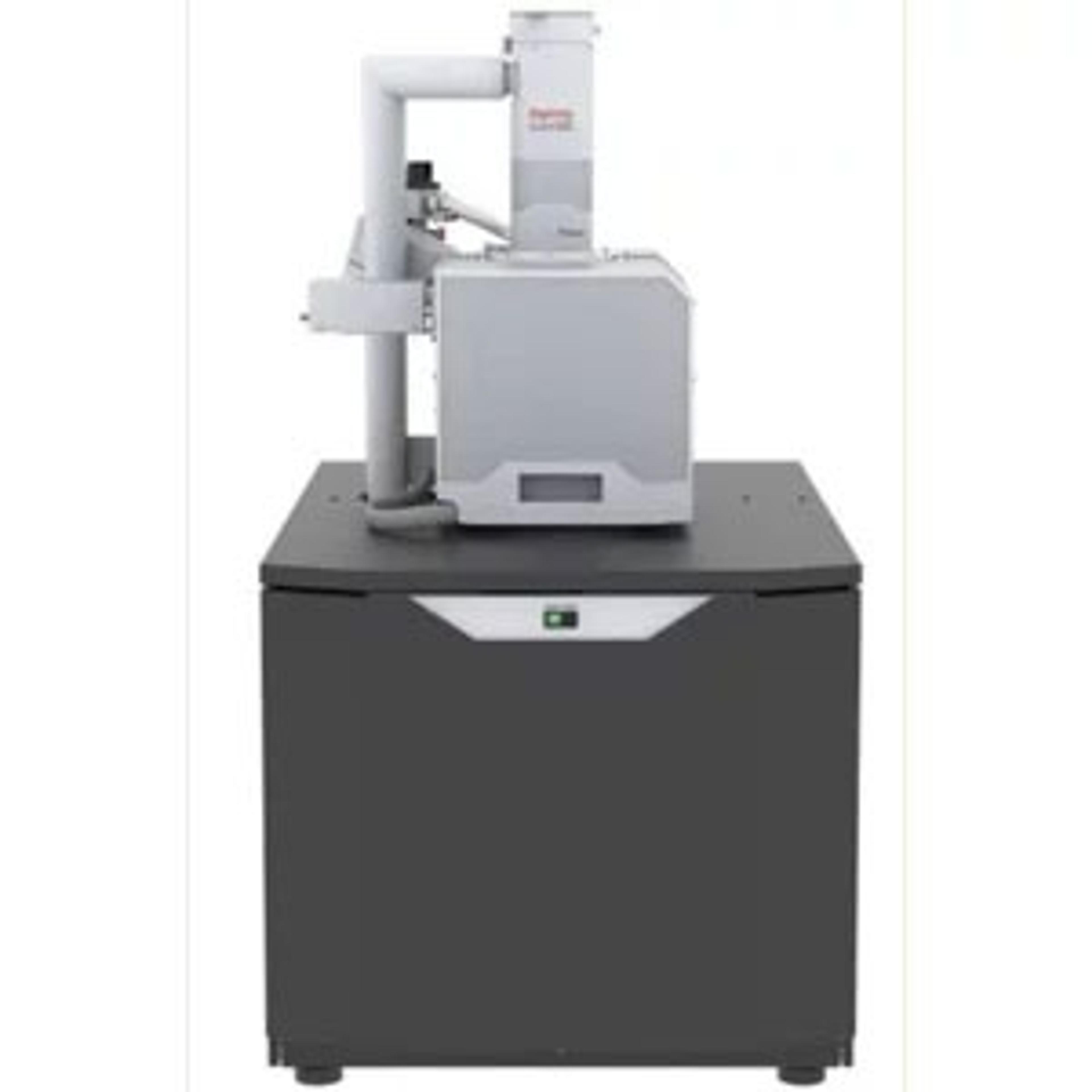
How ColorSEM can overcome the challenges of SEM and EDS analysis
Discover on demand how ColorSEM can provide instantaneous, reliable and complete elemental analyses for material characterization
6 Apr 2020

EDS analysis (energy-dispersive X-ray spectroscopy) is a powerful technique used in conjunction with scanning electron microscopy (SEM) for the study of elemental sample composition. This process is considered relatively slow, running on a separate acquisition system and generating greyscale images.
In this on-demand SelectScience webinar, Brandon Van Leer discusses how Thermo Scientific™ ColorSEM™ Technology could possibly solve these issues by altering greyscale SEM into a color technique with instantaneous elemental information.
Read on for highlights from the live Q&A session or register to watch the webinar at any time that suits you.
Watch Webinar NowQ: What are the detector areas that are needed for ColorSEM?
BVL: ColorSEM is all about user experience, allowing the user to select any detector size. Typically, users choose a 10-square-millimeter detector, but there's no reason why they can’t pick a larger detector. It's a question of how the software handles the acquisition of the X-rays, and how the software quantifies these X-rays to form the final image.
Q: What are the best measuring conditions for high-resolution EDX mapping with a tungsten filament SEM?
BVL: It’s important to be aware that ColorSEM is always switched on. This means that as long as X-rays are being generated, they will be acquired. The signal will be continuously ready and available to the user via the user interface. Regarding what the best conditions are for high-resolution EDS, it comes down to recognizing that X-ray signal generation is a subsurface phenomenon. Ask yourself whether the information you're interested in is sitting at the surface or is it buried information. This is significant, as you're going to be receiving X-rays, not only from the surface, but from those buried features, occasionally convoluting the signal.
In my own experience using SEM and EDS, I often try to remain on the lower end of the scale, from 10 to 15 kV. In some cases, it's possible to go even further down to 3 to 5 kV. This is dependent on the type of system you have, and whether it's windowless or not. Depending on the current and the tungsten filament system, resolution can be quickly lost. Selecting large beam currents will generally generate blurry images, perhaps losing sight of what it is a user is trying to generate information on. I’d say that 1 to 10 nano-lamps is the ideal region to acquire high-resolution EDS.
Q: Does the system notify you when there is a peak overlap?
BVL: No, the system does not notify you when there is a peak overlap. Instead, what we are doing is using quantified analysis by applying NORAN quant algorithm calculations. Therefore, what you are seeing are quantified results, both in the image as well as in the data you collect.
Q: Can Color SEM be used without EDX installed in 7 Pro?
BVP: Currently, ColorSEM is dedicated to our lab-based systems. As a result, ColorSEM on the Prisma E is available, and we'll soon be moving forward to bring this technology to the Quattro, Schottky field emission system.
Q: The elemental identification, is it normally done with X-ray?
BVP: Yes, elemental identification is normally achieved with X-ray. One requirement for this is that you must use an EDX detector. ColorSEM is based on X-ray information, but what's unique about ColorSEM technology is that we capture and process the signal along with the image signal. This allows the user to see a combination of both the secondary electron, the backscatter electron image, as well as the X-ray information that's being collected by the detector.
Q: Is this system available for sale now? Is it possible to get a system to try in the U.K.?
BVL: Yes, this technology is now available. We have a demonstration lab situated in Eindhoven, the Netherlands. We’ve recently introduced live Webex/webinar demos, allowing possible users to see the system in action.
Q: Can this ColorSEM detect all common elements including sulfur?
BVL: Absolutely. Yes.
Q: Does ColorSEM only work with inorganic material?
BVL: No, ColorSEM works with a variety of materials. If a user is trying to gather information on a polymer material, such as plastic, EDS would not be a suitable technique. Instead, FTIR or Raman would be more appropriate. However, if a user is looking to identify potential organic contaminants, then EDS could be the correct technique.
Q: Can you share what additional components are present in the instrument to facilitate ColorSEM compared to a normal SEM?
BVL: The main difference between SEM and ColorSEM is that an EDS detector and a scattered electron microscope are required. In addition, we provide additional hardware for pulse processing and signal generation, as well as a pattern engine generator.
Q: I need a higher current for EDS mapping. How much current is necessary to use ColorSEM mode?
BVL: Whether a user is using two pico-lamps or five nano-lamps, the process is down to the quality of the overall image and the time it takes to generate the image. The lower the beam current, the smaller the number of generated X-rays collected by the detector. This will lead to the process taking longer to generate a high-quality ColorSEM image, since the user is taking those X-rays and generating an image based on the NORAN quantification algorithms. This could be quicker using STEM EDS, where generally the user will be working with a smaller beam current, 150 to 500 pico-lamps. Overall, this is dependent on the sample type, as well as the beam current.
Q: Does the software also work with other SEMs? For example, a ZEISS SEM and an Oxford Instruments EDX?
BVL: At the moment, the software doesn’t work with other SEMs. This is a proprietary technology that is dedicated to Thermo Fisher electron microscopes.
Q: What kind of restrictions does ColorSEM have with respect to the nature of samples, say, in dimensions, magnetics, or other properties that hinder the exhibition of a particular color?
BVL: I would say that any limitations would be making sure the user uses good sample preparation protocols, to ensure the sample is electron microscope ready. If a sample is extremely wet, and the user is not in low vacuum or ESEM mode, vacuum incompatibility will cause issues. However, in terms of working with either a magnetic specimen or even a ceramic, you would still have to apply the same kind of protocol to make sure that that sample can be imaged well inside an electron microscope. Also, if a user overcoats the specimen, then perhaps the signal could be the coating itself and not the sample.
Q: Is quantitative information obtained and how accurate can it be?
BVL: ColorSEM is based on the NORAN quantification algorithm systems with algorithm software. NORAN was one of the first elemental-dispersive spectroscopy manufacturers. In addition, they made some quantification algorithms that have been deemed highly accurate. We have performed many studies investigating these standards in geological specimens and bulk materials, generating errors less than 5%.
SelectScience runs 3-4 webinars a month across various scientific topics, discover more of our upcoming webinars>>