Illuminate Intracellular Calcium Kinetics in Real Time with Live-Cell Imaging
How advanced imaging technology is progressing drug discovery through understanding of GPCR-dependent second-messenger systems
30 Nov 2018

Cell signaling cascades resulting from interactions between G-protein coupled receptors (GPCRs) and their ligands control a wide variety of intracellular processes, making them major targets of drug discovery efforts, particularly for cancer treatment. The cell signaling cascade takes the form of a complex, multifaceted series of second-messenger systems that act in concert. One important second messenger in GPCR-mediated cell signaling is calcium (Ca2+), which plays a critical role in cell response to intracellular and environmental cues. The ability to detect rapid changes in intracellular levels of calcium with high temporal resolution is key in characterizing GPCR activation.
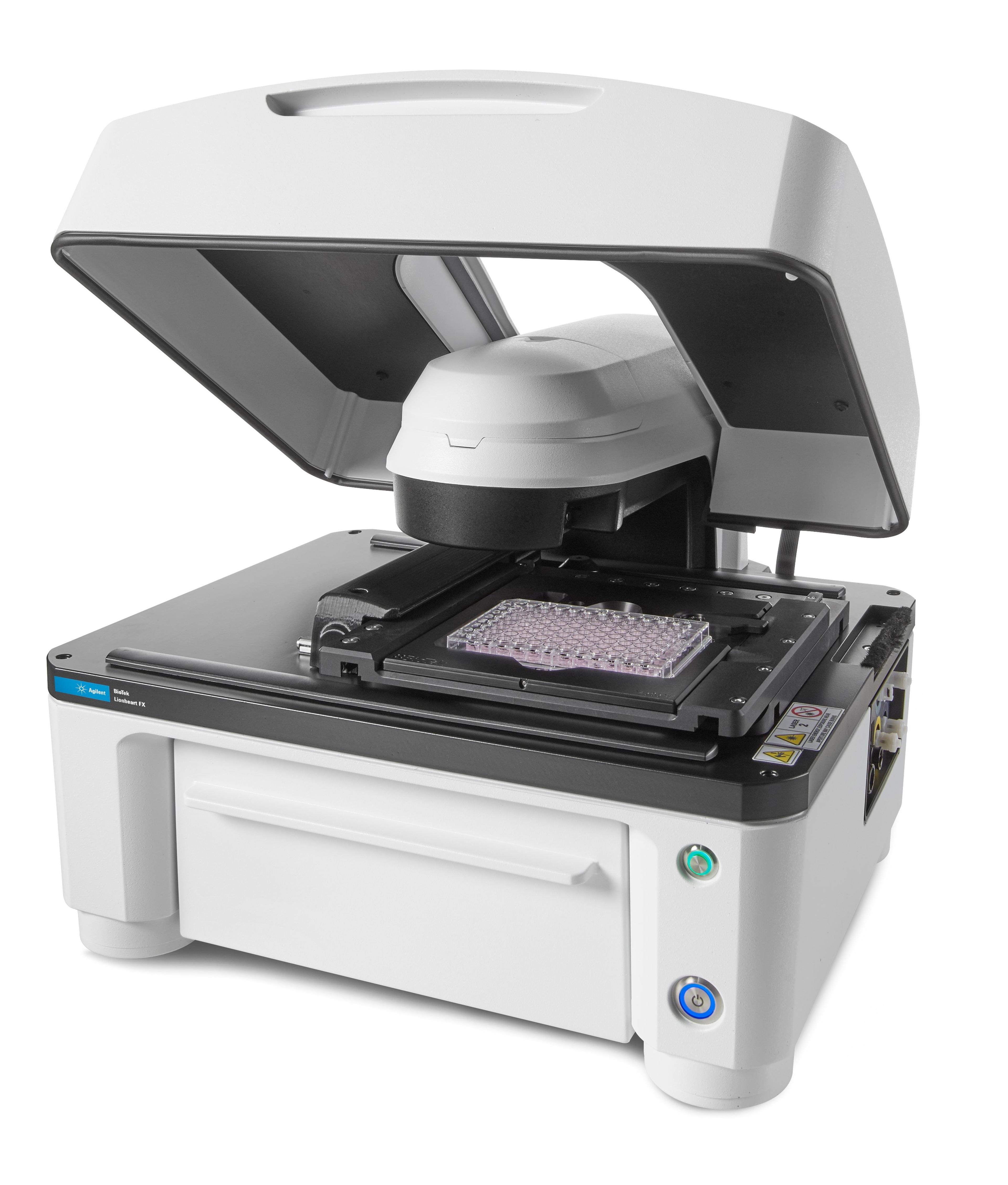
BioTek recently launched the Lionheart™ FX Automated Microscope – a compact, inclusive microscopy system – ideal for the calcium imaging workflow. In this article, SelectScience® speaks with Dr. Peter Banks, Scientific Director of BioTek Instruments, Inc. to learn how the company’s latest cell imaging technology can advance calcium flux analysis, by imaging individual cells with sub-second temporal resolution.
Why is calcium imaging a hot topic?
We are primarily interested in calcium mobilization as it pertains to G protein-coupled receptor (GPCR) activation. This gene family has been and remains one of the most actively pursued for drug discovery. Many different types of instruments have been used to detect calcium mobilization, including microplate readers measuring fluorescence intensity with dyes like Fluo-4. Imaging which enables the visualization of each cell in the field of view, used in conjunction with sophisticated image analysis routines, allows for the segregation of cells that respond fully to GPCR activation, providing greater assay performance than that achieved using an average response of all cells.
Advancing Calcium Imaging
Tell us about some of the features of the Lionheart that have helped to advance calcium imaging?
- Lionheart FX has on-board injectors that allow for uninterrupted kinetic measurement of the rapid onset of calcium mobilization.
- The speed and sensitivity of signal acquisition with Lionheart’s camera provides multiple frames per second capability, necessary for the appropriate time resolution to fully characterize the transient calcium mobilization signal. This can vary widely, depending on choice of GPCR, whether it is endogenously expressed or transfected, cell line and/or agonist.
- Our Gen5 imaging and analysis software provides automated operation from focus and exposure settings to image processing and analysis such that whole microwell plates can be rapidly imaged and analyzed for calcium mobilization.
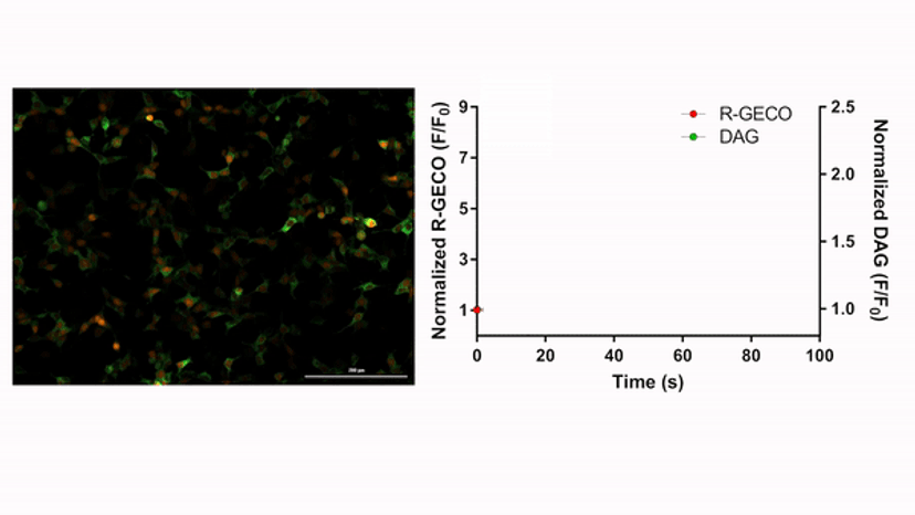
Video: Left: A time-lapse movie of calcium ion (red) and diacyl glycerol (green) production upon addition of an agonist to live cells (left). Multiplexed detection of these second messengers enabled using Montana Molecular’s biosensors. Right: The graph demonstrates quantitative determination of flux, where data production is timed to the time-lapse movie.
Dr. Banks’ 5 Tips for Calcium Imaging and Analysis
Consider using phenol red-free growth medium to minimize background fluorescence. Alternatively, remove media and replace with PBS just prior to running your experiment.
Consider injection volumes that are ~20% of the starting well volume (e.g. 20 mL 6x agonist into a well containing 100 mL). This ratio promotes efficient and rapid mixing with minimal disruption to the cell layer. This is particularly important for cells that are loosely attached to the plate.
Test a couple of wells to determine optimal experiment duration that captures peak calcium release and sufficient return to baseline.
Similarly, use the test wells to confirm an appropriate image capture rate. A lot of GPCR pathways do not require 20 or even 10 frames per second to generate well-populated kinetic curves. More information is not always better, as this requires more processing time.
Exposure settings should result in visible, but faint, pre-stimulated cells, allowing for a large change in sensor fluorescence (dynamic range) upon addition of the agonist.
Customer Feedback
What feedback have you had from scientists about using the Lionheart for calcium mobilization?
- Our customers have reported a much larger assay window when using our automated imaging-based approach compared to conventional methods that rely on bulk fluorescence intensity changes (6 to 8-fold compared to ~2 fold).
- The ability to call up an existing protocol to run additional experiments is convenient and provides reproducible results.
- Customers like the ability to look at the experiment images to confirm results and generate videos for presentations.
- The detailed analysis provides unique insight into the biology (difference in subcellular calcium flux kinetics, percent responders, multiple cycles of calcium flux per cell, etc.) that is lost when measuring bulk fluorescence.
Looking to the Future
What do you see for the future of GPCR signaling pathway analysis and the technologies involved?
GPCRs use multiple pathways for cellular signaling involving a number of second messengers. Being able to simultaneously measure transient responses in live cells from multiple second messengers will provide a much greater understanding of GPCR signaling and this is where I think the future of GPCR analysis is heading. BioTek is currently collaborating with a reagent company called Montana Molecular that provides GPCR biosensors for many GPCR second messengers, including calcium, cyclic-AMP, diacyl glycerol and PIP2. These biosensors are provided with different spectral characteristics that allow for the multiplexed analysis of GPCR second messengers.
How do you hope BioTek technology will advance drug development/treatments for disease?
BioTek’s Automated Cell Imaging product line brings highly flexible, cost-effective cellular imaging to just about any laboratory, whether it’s those in big budgeted Pharma, the more cost-conscious academic lab or spin-off start-up. We hope to enable a wide range of customers interested in better understanding disease onset, progression, diagnosis and treatment.
Resources
Learn more about the cell imaging technology available from BioTek and how it can be used to characterize calcium flux.
- Application note: Characterizing Calcium Mobilization using Kinetic Live Cell Imaging
- Application note: Automated Imaging Assay for Characterizing Ca2+ Flux with R-GECO Biosensor
- Poster: Automated Imaging-Based Technique for Monitoring of Ca2+ and DAG Biosensors Enables Detailed Characterization of GPCR Kinetics