Image registration and spatial infiltration analysis in HALO
Watch this on-demand webinar to learn how an advanced spatial analysis workflow can be used to analyze the infiltration of CD8 positive cells across the tumor-stroma interface
6 Jul 2021
Image analysis is an ever-evolving field of digital pathology that has traditionally been used to perform relatively simple quantitative analysis. Modern analysis approaches allow for increasing amounts of quantitative data to be extracted from histological sections.
In this on-demand webinar, research scientist Andreas Theodosi and image analysis manager James Clay take a look at the application serial section registration of pan-cytokeratin, estrogen receptor, progesterone receptor, and CD8-stained serial sections of human breast carcinoma samples.
We demonstrate how, through tissue classification of pan-cytokeratin stained tumor in one serial section, we can restrict the analysis of estrogen and progesterone receptor positivity to the tumor area in the registered serial sections.
We also demonstrate how an advanced spatial analysis workflow can be used to analyze the infiltration of CD8 positive cells across the tumor-stroma interface
Think you’d benefit from this webinar but missed it? You can now watch it on demand at any time that suits you and read on for highlights from the Q&A session.
Watch on demandQ: Can HALO work with images from a different scanner, specifically, an Aperio?
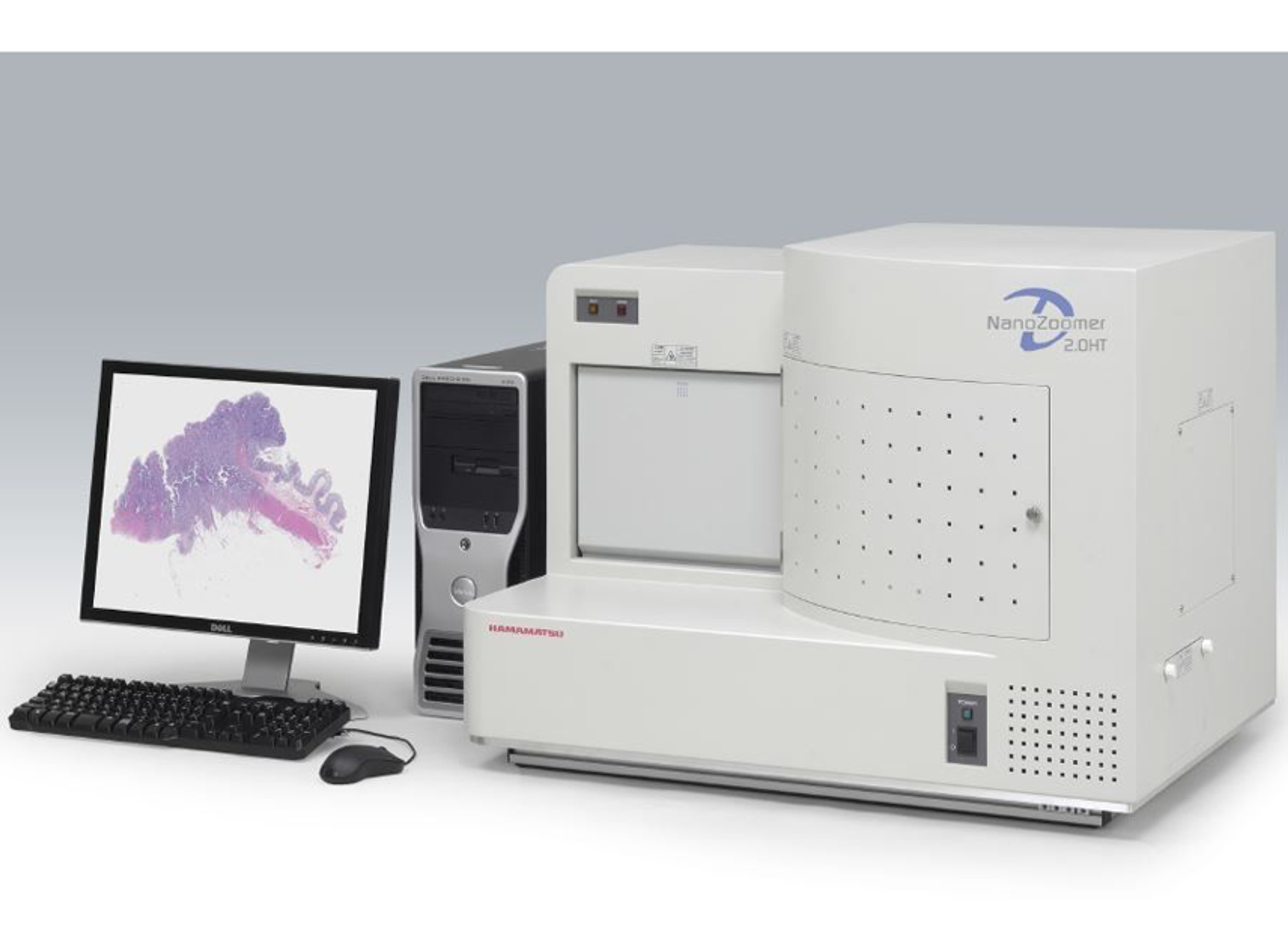
AT: Starting with the Aperio, yes, it can. It can work with both svs and afi file types. It can also work with a whole range of other file types such as jpegs and gifs. It can also work with Leica, Hamamatsu, Nikon, 3DHISTECH, and Olympus Light. It can take images from pretty much any platform.
Q: Can spatial analysis be used with multiplex chromogenic or fluorescent slides?
JC: We can use spatial analysis on either chromogenic or fluorescent stained slides. In the presentation, we used chromogenic over stereo-section registration. There is also the option to do that in spatial analysis on single-section multiplex slides, be it chromogenic or fluorescence. There are benefits to taking either approach, whether it be chromogenic or fluorescence.
In chromogenic assays, we can sometimes struggle with the convolution of the two stained chromogens. If we see the signal in the same cell populations and the same cell compartments, it can be very challenging to separate the two signals and get a reliable detection. In those scenarios, it is common to use fluorescence multiplex to reduce where you can separate the fluorophores from staining into separate channels, which are analyzed as discrete entities. You can get a clearer cell detection and analysis using fluorescence. Histologically, we have both bright field and fluorescence imaging capabilities, so we can support our clients with either approach.
Q: At the moment, we do IHC and ask a pathologist to assess our stained research samples. I like the advantages and extra data that you get with digital image analysis. Do you work with pathologists to identify tissue regions of interest in human tumors, for example?
JC: Yes, we can certainly take that approach and we agree that a lot of the pathologist's reads are very useful and required in many studies. There is also a lot more quantitative and granular data that we can extract from imaging the same slides using image analysis. Where we do require very specific annotation of human region of interest, or for example in the staging and grading of different neoplastic lesions, we work with both medical and veterinary pathologists to mark the images. Then from the source and digital scan, we can pull those annotations into HALO and use those as the basis for the quantitative analysis of the region of interest.
Q: How reliable is image analysis? Why would you use it instead of manually reporting on slides?
AT: It is incredibly reliable. Once it has been tuned and it is being tuned by an operator who's got experience with the platforms, you can tune up an accurate algorithm and that it will always produce that reliable output. It removes operator bias once the algorithm is tuned. Would I use it instead of manual reading the slides? I think so, but as James alluded to, there are going to be some situations where it is going to be mandatory for the study output. We can work synergistically with the pathologists and help them guide the processes if required.
Q: How have you made your choice between weak, moderate, and strong staining?
JC: There are various approaches we use to set our intensity gates and stain intensity thresholds in the algorithms. In this instance, with the issue with progesterone, we use non-positive controls that pathology scored grade one plus two and three plus samples and use those as the basis to set the boundaries for the weak, moderate, and strong staining.
In other study designs, it depends on the nature of the work involved and the requirements of the clients. Common approaches would be to use over-expressing and under-expressing wild type transfecting cell lines to help set those intensity gates. Known pathology scored case samples, and again, it's the kind of area that, where required, we can work with the study pathologist to help set those gates.
Learn how an advanced spatial analysis workflow can be used to analyze the infiltration of CD8 positive cells across the tumor-stroma interface>>


