Imaging Cell Ultra-Structure Using 3D Correlative Microscopy
SelectScience® spoke to Dr Louise Hughes, Bio-Imaging Unit, Oxford Brookes University, about the technology her lab cannot do without
29 May 2016

SelectScience® spoke to Dr Louise Hughes, Bio-Imaging Unit, Oxford Brookes University, about the technology her lab cannot do without
European Molecular Biology Laboratory (EMBL) Founded in 1974, EMBL is Europe’s flagship laboratory for the life sciences – an intergovernmental organization with more than 80 independent research groups covering the spectrum of molecular biology.
The Bioimaging Unit at the Department of Biological & Biomedical Sciences, Oxford Brookes University, has access to one of the most comprehensively equipped biological microscopy suites and offers a range of services to students, staff and outside clients. At the workshop ‘From 3D Light to 3D Electron Microscopy’ (EMBL 3D), jointly organized by the European Molecular Biology Laboratory (EMBL) and ZEISS Microscopy, SelectScience spoke to the Unit’s manager, Dr Louise Hughes, about her research and the innovative technology she uses.
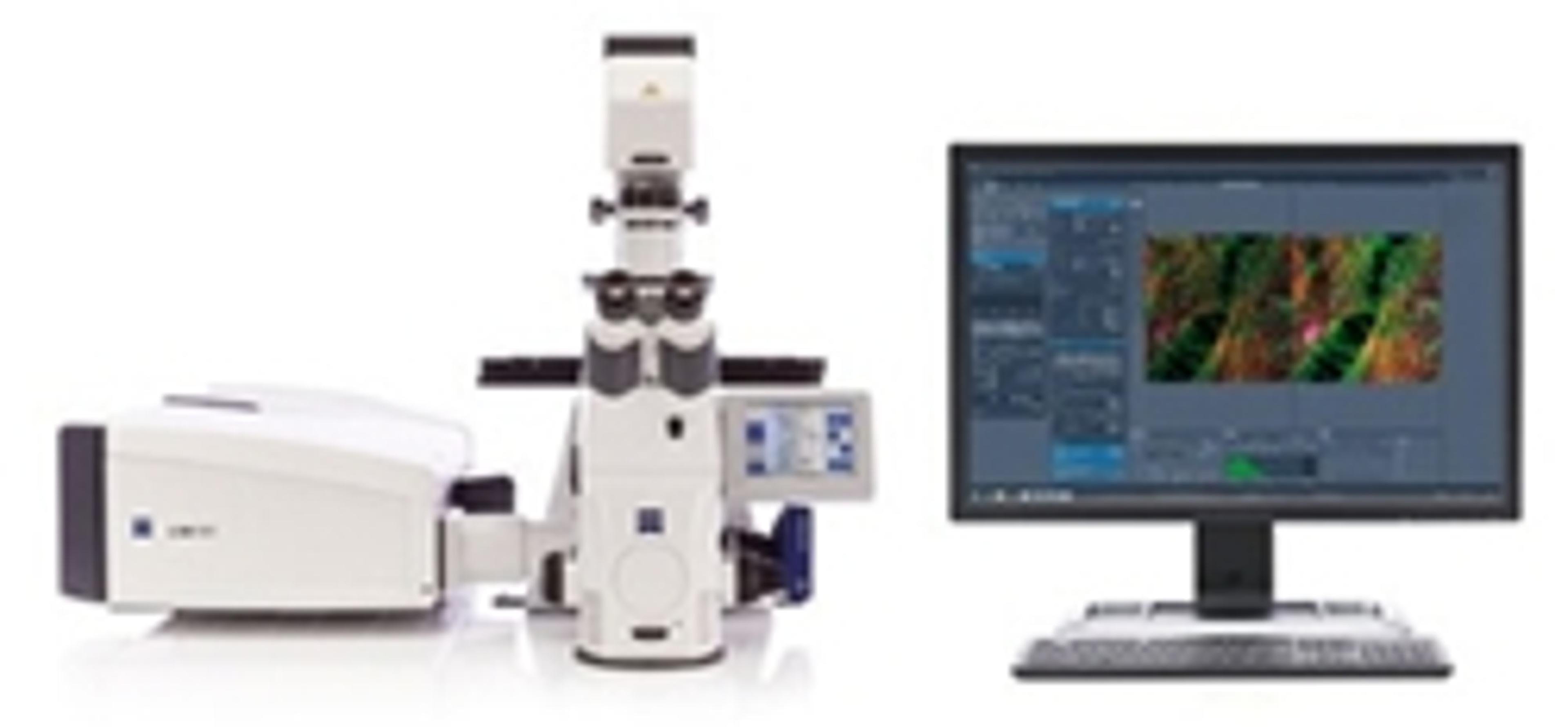
The lab “predominantly does plant research”, explained Dr Hughes. “We also research into parasites and viruses, and general cell ultra-structure.” There, researchers are looking at “how organelles are moving within the cells and at various constructs that we’ve created within cells” using a ZEISS LSM 880 with Airyscan that’s “got everyone ridiculously excited”, Dr Hughes revealed.
These “better resolution techniques” allow scientist to look in more depth at the specific biological processes they are studying. With three dimensional (3D) electron microscopy, “we are able to get really deep inside tissues and look at how organelles are organized throughout different cellular structures”, whether in plants, parasites or animal tissue. For example, Dr Hughes has been looking at organelle and cellular changes over the whole cell cycle “if we’ve introduced some genetic mutation, or whether they’ve been grown in different environments”. Although traditionally it has been difficult to interpret three dimensions from 2D images, “3D imaging has really changed the way we look at biology and how cells function”.
The future of biological science
Although electron microscopy is “fantastic and the resolution amazing, you are always looking at an artificial situation because everything is dead and fixed and immobile”, Dr Hughes explained. Light microscopy gives you “the ability to look at the dynamics of what’s going on at the cellular and tissue level”, she continued, “but you don’t have the resolution”. The Oxford Brookes unit currently uses both light and electron microscopy, but not on the same sample – “it’s important for us to get to the point where you take one sample all the way through, from live imaging looking at the fluorescent markers through to the EM level looking at three dimensions with electron microscopy”, said Dr Hughes.
The technology is at “the very early stages, but the impact that correlative microscopy is going to have is unquantifiable”, Dr Hughes explained. “The ability to integrate light and electron microscopy is so key to biological processes that we can’t put a limit on where it goes…the sky is the limit.”
Find out more about the other speakers and presentations from EMBL 3D, or learn more about the applications of Correlative Microscopy.
Watch our interview with Dr Hughes here.


