Improve and Automate Your Western Blots
Learn about modern immunoblot imaging technologies that can improve your data quality and save you time
23 Sept 2018
When you plan, set up and perform your western blots, there’s always one vision for the final outcome – the appearance of those crisp bands. With so many variables at different steps of a western blot process, more and more efforts are being made to automate and eliminate errors. For example, pre-made buffers, pre-cast gels, and dry/semi-dry transfers are all designed to make the western blot process easier. Today we speak with Brian Webb, senior R&D manager at Thermo Fisher Scientific, to learn about technologies for the final step of your western blot – image capture and analysis. Read on to learn how this technology can reduce hands-on time and yield reliable data.
Please introduce yourself and your role at Thermo Fisher
My name is Brian Webb. I am a senior R&D manager at Thermo Fisher Scientific located in Rockford, Illinois. My R&D group was responsible for developing the Invitrogen™ iBright™ Imaging Systems platform.
Can you expand on the history of the iBright? Why and how was it developed?
My team has developed many instruments and reagents for the western blotting workflow over the past 10 years. As we observed customers perform the final and most important step of the western blot process – the image capture and analysis step – we realized there was an opportunity to improve the user experience and throughput by designing a new imaging instrument with new features to streamline and automate this process.
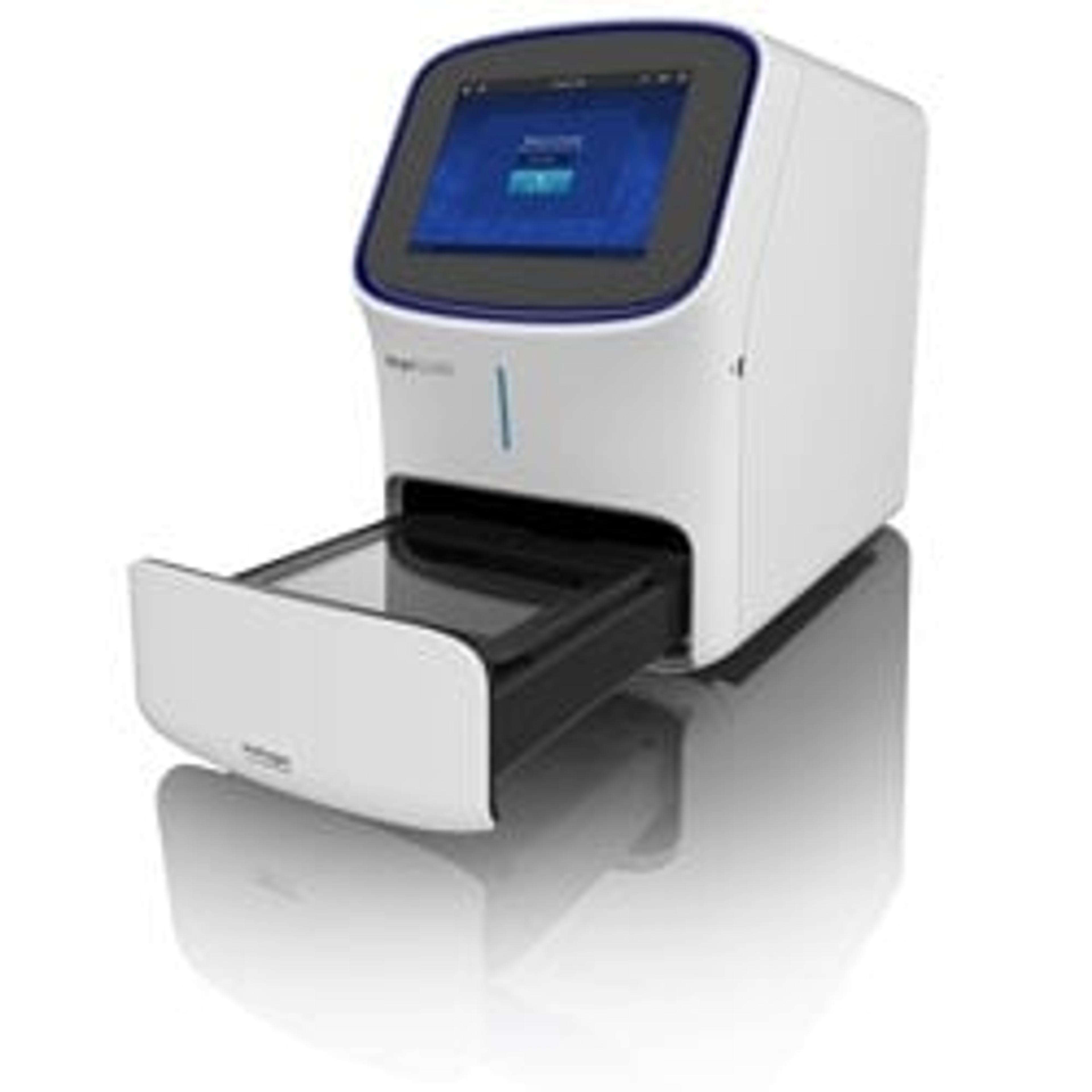
It has been a lot of fun work with our team of scientists and engineers to take our customer experience “wish list” and make them a reality with our iBright Imaging Systems. The iBright platform is truly unique and I am confident it will delight our customers. The platform consists of the iBright CL1000 Imaging System for capturing signals from chemiluminescent western blots, stained protein and DNA gels. The iBright FL1000 Imaging System is our premium offering and expands upon the capabilities of the CL1000 model, with added capacity for fluorescent western blot imaging.
What are the key benefits of the iBright compared to the other imaging systems?
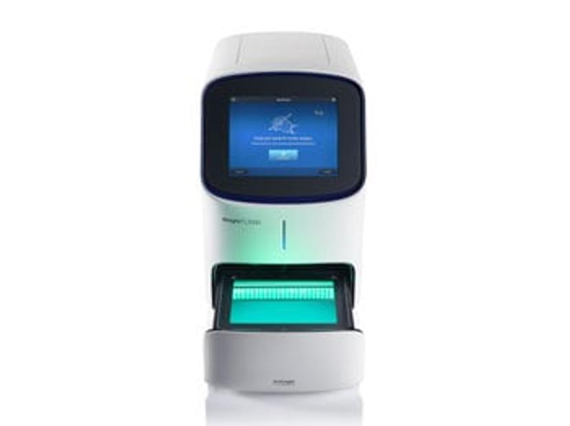
We designed iBright Imaging Systems with the customer in mind and a goal to obtain optimal western blotting results with minimal hands-on time. Many steps of the western blot image capture process that require manual inputs on other suppliers’ instruments have been automated on the iBright Imaging Systems, including sample alignment, focus and zoom. You can simply place your blot on the transilluminator tray and the system does the rest.
The next key decision in western blot imaging is deciding on an optimal exposure time that provides the maximum dynamic range without saturated pixels. Our patented Smart Exposure™ technology utilizes a sophisticated algorithm to automatically calculate the optimal exposure time with a single click. After image capture, the iBright Imaging Systems have an automatic lane and band finding feature, for effortless and accurate sample analysis. The large capacitive LCD touch screen is designed with responsiveness to inputs like pinch and zoom, and panning that enables you to navigate your images and data quickly and efficiently.
We also designed this platform with safety and the environment in mind. The iBright Imaging Systems use long-life green LEDs instead of UV bulbs in the transilluminator, eliminating your exposure to UV rays and eliminating mercury waste generated from UV bulbs. Finally, to address the growing need to access data and perform analysis on the go, we developed the first cloud-based western blot image analysis software on the market. These features help you worry less about the tedious aspects of western blot imaging and focus more on your data.
What are some unique application areas for the iBright? What can scientists do now that they couldn’t do before?
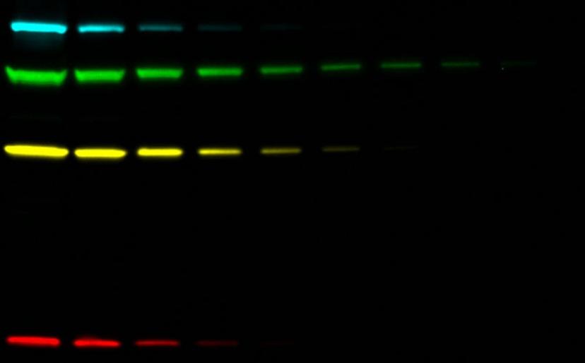
The iBright FL1000 Imaging System offers five fluorescent channels, spanning both the visible and near-IR range. This enables up to four-channel fluorescent multiplexed western blots, so users can visualize proteins of interest in different states, multiple proteins in a pathway, or load control proteins on the same blot at the same time. Our large transilluminator, with a 22.5 x 18 cm field of view, enables the capture of up to four mini blots or gels at a time. The advanced on-board analysis software we developed provides automatic lane and band finding that actually works.
Band quantitation and densitometry can be performed in a fraction of the time, helping prevent a backlog of users waiting to use the instrument. Our on-board analysis software provides the ability to analyze four independent blots or gels within the imaging area, all at the same time. This feature, combined with the ability to capture up to 4-plex western blots, expands the potential to study multiple proteins and experimental conditions during an imaging session.
What do you see for the future of this product? And what is the next big step for gel doc imagers?
Capturing western blot data with film is progressively becoming a thing of the past, as imaging instrumentation becomes more widely available. These instruments are continuously evolving to meet the needs of scientists. We see iBright Imaging Systems becoming an integral component of the western blotting workflow and we take feedback from researchers very seriously and make every effort to incorporate that feedback into our instrumentation design and development. The core ethos of our western blotting devices and instrumentation is to strive to be at the cutting edge, while also being accessible and user-friendly.
Do you use the iBright Imaging Systems in your lab? We’d love to hear from you. Please leave a 2-minute review here.