Improving acquisition and analysis of 3D cell model assays with water immersion objectives
Sebastian Peck discusses an automated high-content imaging solution designed to deliver more precise results from complex biological assays
13 Sept 2020

Sebastian Peck is a senior product manager for cellular imaging at Molecular Devices, one of the world's leading providers of high-performance bioanalytical measurement systems, software, and consumables for life science research, pharmaceutical, and biotherapeutic development. Peck received his bachelor’s degree in microbial biology from University of California, Berkeley, and has over 10 years of experience in imaging, high-content screening, and life science technology. Currently, Peck specializes in maximizing high-content imaging solutions to best meet the scientific community’s needs.
Here, Peck discusses Molecular Devices' innovative water immersion imaging technology with SelectScience®. This technology helps researchers conduct high-content screens of complex three-dimensional (3D) models, delivering crisp images while optimizing assay sensitivity, flexibility, and throughput.
Expanding our knowledge in the life sciences will rely heavily on research using human-derived 3D models. What are the challenges that arise when working with such complex systems?
Given their simplicity to handle as well as to image, two-dimensional (2D) monolayers remain the standard for drug discovery and screening. However, researchers are increasingly moving toward three dimensional (3D) models such as organoids, spheroids, and organs-on-a-chip to obtain more physiologically relevant data. This transition presents a few challenges for researchers, with image quality topping the list. The thicker the 3D sample, the more difficult it is to get clear images due to lack of optical penetration and increased aberrations, which ultimately leads to limited results.
Molecular Devices develops 3D imaging products that are empowering scientists to achieve new discoveries. Tell us about your technology and mission.
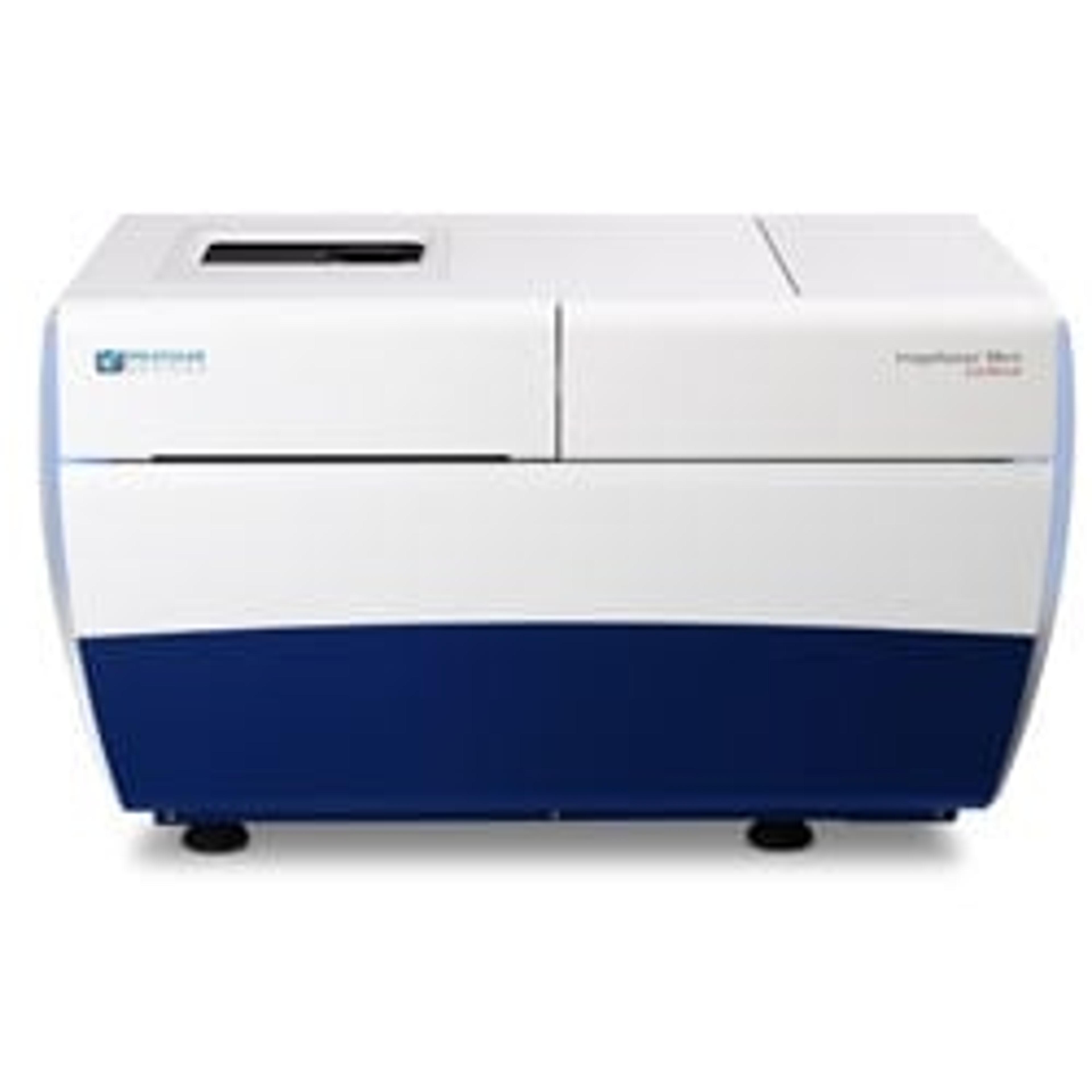
Leveraging 37 years of industry experience, our life science solutions help fast-track scientific discoveries to improve quality of life everywhere. This year, we introduced a new automated water immersion imaging technology for the ImageXpress® Micro Confocal High-Content Imaging System, which builds on the company’s existing 3D imaging capabilities. The high-performance water immersion objectives facilitate the growing demands of 3D imaging by enhancing the resolution, sensitivity, and throughput to improve acquisition and analysis of a variety of complex cell-based assays.
What capabilities does water immersion technology enable for drug screening?
Imagine standing at the deep end of a swimming pool and glancing down into the water with the naked eye at dive rings piled together on the floor. The rings are blurry, right? Put a pair of goggles on, press them to the surface of the water, and individual rings appear clearer having removed much of the air between the eyes and the targets. Water immersion technology is similar in that it provides water as a conduit for the light path between the objective and the sample. Because the water closely matches the refractive index of the sample, it decreases aberrations when imaging deep into 3D models. In addition, it allows for a higher numerical aperture, as compared to air, which can improve lateral and axial resolution, as well as allow up to four times better light collection. This improved clarity offers insights that might otherwise be missed or overlooked using lower resolution imaging technology.
The automated, hands-free nature of the ImageXpress Micro Confocal system also gives researchers the confidence to acquire data in high-content format, unattended. The water immersion system automatically adds and removes water for continuous uptime with unparalleled resolution and speed, reducing acquisition time by up to 30 percent in a two-color assay (384-well plate). This is a significant benefit to both 3D and 2D screening.
What are researchers able to see with water immersion objectives for automated high-content imaging?
In addition to the benefits for 3D assays, water immersion also improves 2D imaging of subcellular details such as mitochondria. Our scientists recently monitored mitochondria dynamics and their phenotypes with high-content imaging using water immersion objectives. The combination allowed them to effectively determine not only mitochondria count (they were able to see between 24 and 34 percent improvement in Z’ values for the number of mitochondria – including fused vs. fragmented – when compared to air objectives in this study), but also mitochondria shape which is important for assessing toxicity.
Whether researchers are imaging 2D or 3D models, our innovative water immersion technology offers better visualization of changes in cellular and subcellular components, as well as morphology; more efficient and precise quantification and measurement of phenotypic changes within the cell; and an increased understanding of disease and compound toxicity in cell models.
What does the future hold for 3D imaging and how does Molecular Devices play a role?
3D imaging joins a new generation of technologies that are enabling researchers to speed discovery with precision, accuracy, and individualization. Our focus is to deliver robust, integrated, and easy-to-use life science platforms that enable researchers worldwide to accelerate their work rather than get bogged down by substandard imaging solutions. With a seasoned research and development team and a culture of continuous improvement, we’re committed to pushing the boundaries of 3D imaging with our cutting-edge technologies that advance scientific discovery and solve some of today’s most pressing problems.
For a deeper dive into Molecular Devices' water immersion technology, watch their innovation showcase video here.