Incubation monitoring for cancer cell line quality control and banking
Find out how experts at the Ellison Institute are harnessing new cell monitoring technology to build cancer cell biobanks and enhance cell line quality control and authentication
13 Oct 2020
It is estimated that 1 in 3 people will develop one of the many forms of cancer within their lifetime. Depending upon the type, many will face lengthy bouts of chemotherapy with mixed results. The phenomenon of chemoresistance in some cancer cells is well-documented but not so well understood, and numerous labs across the globe are working on understanding tumor microenvironments and the crosstalk between different cells that is pivotal to tumor progression and response to therapy. Underpinning all this research is the need for quality-controlled, reproducible cell lines, from cancer patient samples, curated in centralized master cell banks.
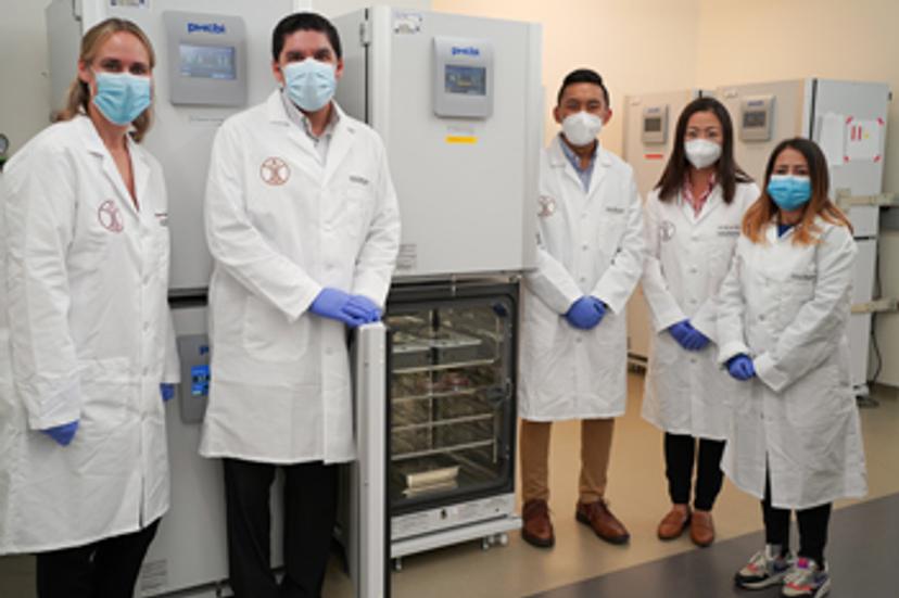
One academic lab at the University of Southern California (USC) in the US is doing just that and harnessing the latest cell incubation monitoring systems to build biobanks with rigorous quality control of every cell line. Based out of the Ellison Institute for Transformative Medicine, we speak to Dr. Michael Doche, Cell Line Team manager, and Dr. Shannon Mumenthaler, assistant professor of medicine and biomedical engineering, to find out what it takes to establish and maintain a high-quality controlled cell line stock for both institute researchers and external collaborators.
Cell banking: The importance of trust
Doche recalls the reasons why the Ellison Institute realized the need to create its Cell Line Team, explaining that in the past they had run into issues with microbial contamination, misidentification of cell lines and researchers sharing cell lines back and forth without really knowing their true provenance and history. “In establishing our workflows and maintaining this cell line bank, the Cell Line Team provides that QC that researchers can count on,” Doche says. “The cell lines they’re re-attaining from us are exactly what they are supposed to be, free of contamination and they have metadata that can be referred back to,” he adds.
Mumenthaler reinforces that message in the context of patient samples, adding: “For patients who have so generously contributed their tumor samples, we make sure that the cellular models derived from their tissue are being used properly and that we’re collecting all the associated metadata so that research projects can easily reference the information.”
Research needs to be reproducible and Doche emphasizes that, for efficiency and reliability, it is important to have every step quality controlled and “ensure that everything is of the highest quality and being conducted in a way that people can trust.”
Achieving that requires an appreciation of the challenges associated with monitoring cell lines and an understanding of the technology that helps overcome those challenges. With collaboration at the heart of scientific advancement, the team at the Ellison Institute has teamed up with Olympus to help them achieve their research goals.
Real-time cell incubation monitoring
The Cell Line Team at the Ellison Institute works with a variety of cell lines, including epithelial-like, fibroblast-like or patient-derived organoids. It is essential to be able to monitor cell lines continuously to remove guesswork over when lines are ready to be split or how well they have been growing overnight or during a weekend. “I can check in real time and look at their history and compare one passage to the previous passage, or one thawed cell line to how it was previously thawed before being frozen again,” says Doche. “It gives reassurance that there are recorded images and data to refer back to,” he adds.
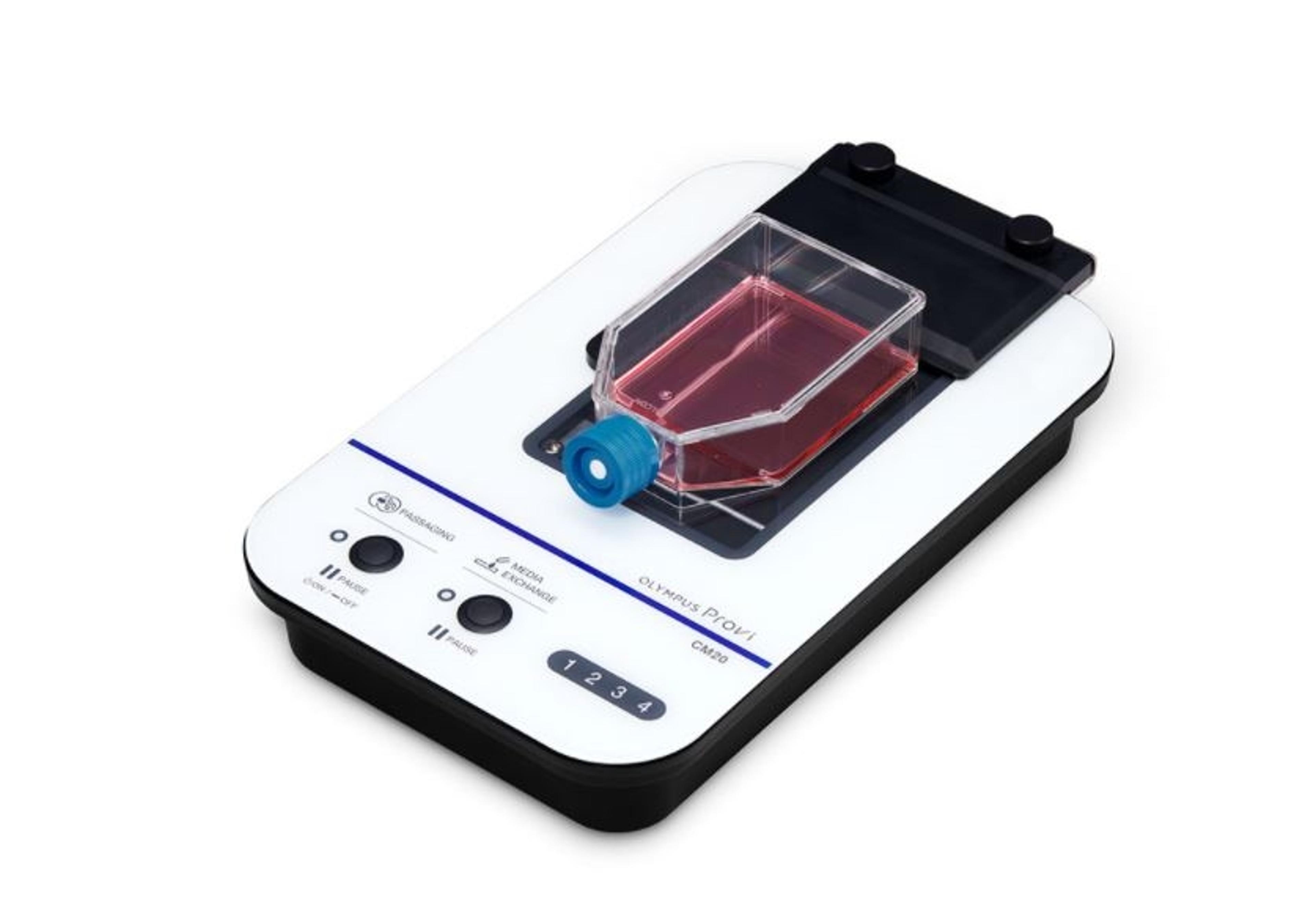
Doche and his expert team use the Olympus Provi real-time cell incubation monitoring system, hooked up to a workstation via either internal wi-fi or, more commonly, via the TeamViewer application which allows remote access from anywhere. “From there, I can access the Provi software directly, and that’s my gateway to seeing the cells whenever I want,” he enthuses. As Doche points out, cells have no concept of weekends or holidays: “I can know exactly when the cells need attention and when to plan a calendar for my team that we can work around.”
Mumenthaler agrees, explaining that they have had far fewer issues with when cells should be passaged or fed, compared with their previous approach to monitoring in person more frequently on site. The Olympus Provi has become essential to their workflow, tracking the health of individual cultures and comparing with historical data to ensure that health is consistent across individuals, experiments and research projects.
A multifaceted partnership
Mumenthaler speaks highly of the partnership with Olympus: “It has helped us with many different research angles that we’re approaching in cancer,” she says. “The Provi system has expanded the kind of data that we’ve been able to collect, and the quantitative metrics we can assign to each cell line,” she further adds.
The Cell Line Team is a very diverse group with many workflows and needs, some of which have been shared with Olympus to potentially add on. “What’s really great about the collaboration with Olympus is there’s a lot of discussion back and forth on the goals that we’re trying to achieve as a cancer research institute and how our partnership is able to help us reach those goals,” Mumenthaler says.
Addressing the bigger picture, Mumenthaler asserts that “we have a very large biobank of samples, from immortalized cell lines to primary patient samples. Being able to have this wonderful biobank with associated metadata has supported a multitude of research projects here at the Ellison Institute.”
Future developments
Looking ahead, Doche predicts that the Ellison Institute’s cell line work will lead to more discovery screening or preclinical screening for either new cancer drugs or repurposed drugs. Doche and his team look forward to “being able to do that in a more automated way with a system that leads us to generate new hypotheses more quickly.”
Mumenthaler talks of coupling a reproducible cell biobank with microscopy tools: “Being able to monitor cancer cells and then measure phenotypic information, such as growth rates and morphology features, in a robust and quantitative manner is very important in the cancer research space,” she concludes.
Do you use Olympus products in your lab? Write a review today for your chance to win a $400 Amazon gift card>>