Integrated Imager Automates Cell Confluence and Growth Monitoring
8 Jan 2010Hamilton Robotics introduces a new Cell Imager for automated cell growth monitoring in the company’s CellHOST culture system. The Cell Imager accurately measures cell confluence and growth in 96-well plates and is ideal for measuring hybridoma clones in antibody screening for therapeutic and diagnostic research and development.
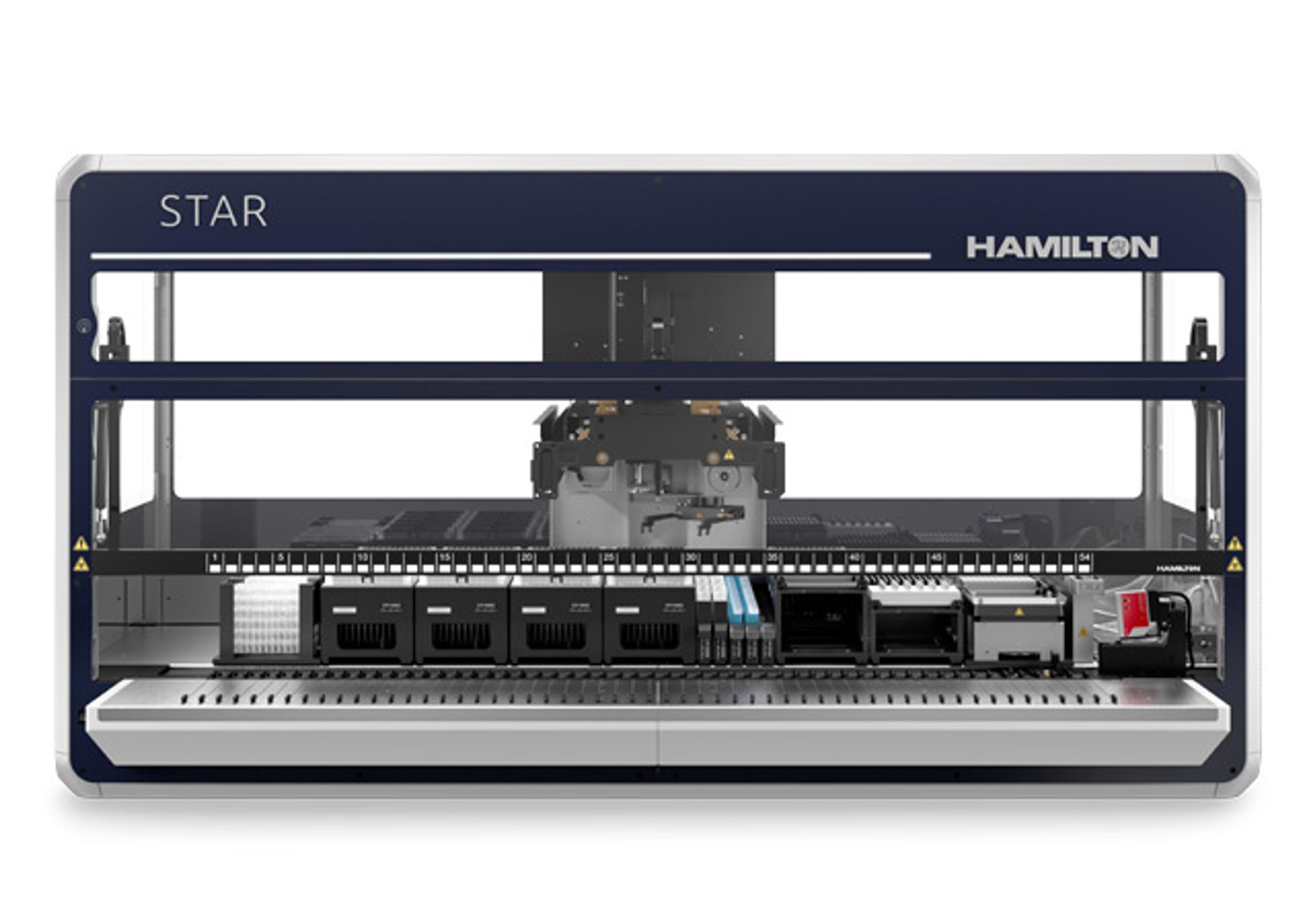
This dedicated imaging module optimizes the cell culture workflow and process by providing timely information with which to make decisions on media changes and subculture steps. The compact Cell Imager can be placed directly on the MICROLAB® STAR workstation deck, and imaging and pipetting are done in the same position for maximum space economy. The design of the Cell Imager eliminates the need for a gripper, which is required for movement of plates to and from other systems on the market.
The Hamilton Cell Imager reads an entire 96-well plate in approximately five minutes. Two positions allow one plate to be imaged while another is processed. The Cell Imager’s software provides live data tracking for a simultaneous overview of cell confluence in all plate wells. The software is easy to use and provides graphical data presentation. It may also be integrated with Hamilton’s Venus Software.
“Our integrated Cell Imager allows researchers to process cells on demand, rather than on a set schedule,” explained Daniel Caminada, Ph.D., cell biology product manager for Hamilton. “This instrument is optimized for hybridoma cell selection and is the fastest and most economical tool for this application.”