No More Western Blotting Worries: Bio-Rad’s New V3 Western Workflow™ Provides Confidence at Every Step of the Immunoblotting Workflow
16 Apr 2016Bio-Rad Laboratories, Inc. launched its V3 Western Workflow, a portfolio of products designed to reassure researchers at every step throughout the immunoblotting workflow. Leveraging stain-free imaging technology, the V3 Western Workflow enables scientists to visualize protein separation, verify protein transfer, and confidently validate blot data via total protein normalization.
The V3 Western Workflow also cuts the traditional western blotting process from two days to one day. From protein separation to protein transfer to load control normalization for quantitative results, Bio-Rad products dramatically shorten time to results while maintaining high performance at each step.
“Instead of two days to do a western blotting experiment from beginning to end, we can see results within a day with the new workflow,” said Dr. Aldrin Gomes, assistant professor at the University of California, Davis.
The V3 Western Workflow consists of five steps, each enabled by Bio-Rad’s family of blotting instruments and/or products:
Five Steps From Separation to Results
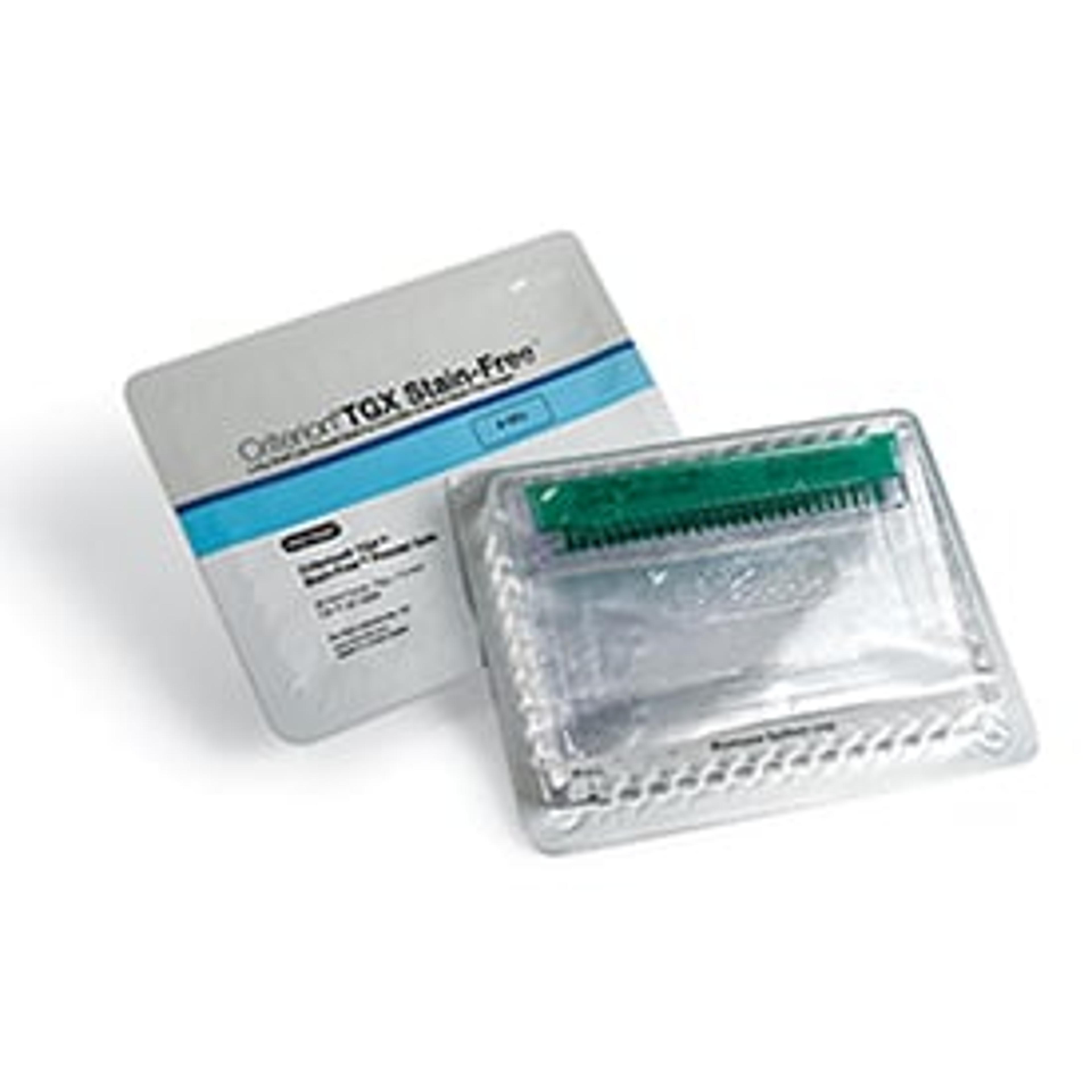
1. Protein separation using Criterion™ TGX Stain-Free™ and Mini-PROTEAN® TGX Stain-Free precast gels
2. In-gel protein visualization using ChemiDoc™ MP imaging system
3. Protein transfer to membrane using TransBlot® Turbo™ transfer system
4. Transfer verification using ChemiDoc MP imaging system
5. Western blot validation using ChemiDoc MP imaging system
Steps 1 and 2: Protein Electrophoresis and Visualization of Separation
Bio-Rad’s Criterion TGX Stain-Free and Mini-PROTEAN TGX Stain-Free precast gels provide researchers with complete electrophoretic separation of protein samples in as little as 15 minutes. The gels include a unique trihalo compound that allows fluorescent detection of proteins in gels or on membranes in one minute when using the stain-free enabled ChemiDoc MP imaging system. Coomassie gel staining methods can take up to two hours to complete.
Steps 3 and 4: Protein Transfer to Membrane and Verification of Transfer
The Trans-Blot Turbo transfer system facilitates efficient protein transfer across a wide range of molecular weights in as little as three minutes with Bio-Rad’s TGX Stain-Free precast gels. Using the ChemiDoc MP imaging system, researchers can instantly visualize protein transfer prior to blot detection by imaging the membrane. The gel can also be reimaged to establish whether any proteins remain.
“We used the ChemiDoc MP to make sure our gels were transferred without any problems,” said Gomes. “It’s less tedious than Ponceau staining. By doing this, we can proceed with confidence knowing we’re not wasting any antibody.”
Step 5: Validation of Western Blot
Researchers quantifying western blot experiments traditionally normalize data with housekeeping proteins, which often requires stripping and probing the membrane a second time, or probing with multiple antibodies simultaneously. Both add extra steps to the protocol, consuming reagents, time, and labor. Furthermore, normalizing against housekeeping proteins might produce inaccurate results.
Using the stain-free detection option of the ChemiDoc MP system, researchers can quantify total protein for normalization purposes. This stain-free total protein normalization method produces faster quantitative results compared with relative normalization results using housekeeping proteins, which can lead to errors.
For more information on how the V3 Western Workflow can increase your rate of success for western blotting experiments, visit the company article page.

