Pioneering researcher creates unique 3D cell culture models to combat liver cancer
Dr. Carlemi Calitz shares her breakthrough in hepatocellular carcinoma research and how it could lead to improved therapeutics and reveals her remarkable finding in cancer metastasis
27 Sept 2020

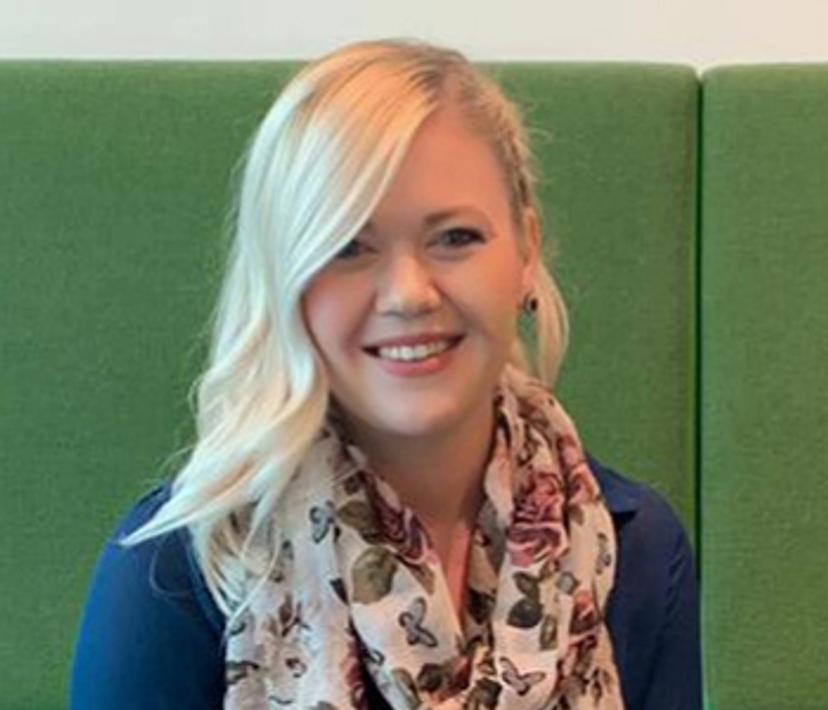
In this exclusive interview, we speak with Dr. Carlemi Calitz, a postdoctoral researcher at Uppsala University working in the group of Dr. Femke Heindryckx, to find out how their team is developing state-of-the-art, three-dimensional models in a bid to tackle hepatocellular carcinoma. We also learn how Calitz accidentally discovered the presence of metastasis by observing “climbing” cells, and how Calitz hopes her models can be used to help others investigate many forms of cancer to improve therapeutics and make her mark in cancer research.
Life in 3D
Hepatocellular carcinoma (HCC) is a primary malignancy of the liver, often affecting individuals suffering from underlying chronic liver disease and cirrhosis. Liver cancer is the second-leading cancer worldwide and is commonly diagnosed at a late stage, when there are little to no treatment options available. Calitz and the Heindryckx group are working to develop a realistic 3D model that scientists can use to help predict drug responses and investigate this and other forms of cancer.
Calitz is confronting how we can better treat HCC head-on by investigating sinusoids. Sinusoids together with hepatocytes are the most basic structure of the liver. “The sinusoid is structured in a way that we have a layer of endothelial cells. Underneath this layer, we then find stellate cells and hepatocytes,” explains Calitz. “Once the liver becomes damaged, the stellate cells migrate towards the hepatocytes and start secreting collagen, type I. This process causes the liver to become stiff, leading to fibrosis, cirrhosis, and ultimately HCC.”
Stepping into a new dimension: From 2D to 3D
The team at Uppsala University uses cell culture plates to help them visually observe endothelial layers on the top of the sample, with stellate cells and hepatocytes embedded into physiologically relevant hydrogel. Calitz explains: “The gel comprising of stellate cells and hepatocytes, is composed of collagen and fibrinogen, causing the stiffness to vary. As a result, we then have a condition where we can mimic fibrosis, cirrhosis and complete HCC.” Calitz developed her unique 3D model based on these components, whereby she was able to observe the three different conditions in one plated system. “If you just focus on targeting one cell and neglect the others, you don't get the bigger picture of what's really happening. That is why we are targeting multiple cells at the same time to get a true feel of what the reaction to a drug will be.”
3D cell culture techniques have continued to receive much attention, since these might provide more accurate tissue models. There is no doubt that 2D and 3D cell cultures are associated with their own benefits and setbacks, with many scientists questioning whether it is now time to start transitioning from 2D to complete 3D cell culture techniques. Calitz explains that her model simply could not be developed using a 2D culture: “It can take seven months to get a mouse model ready to do a study. My model only takes three weeks and can last a while. The model I have in the lab right now is 25 days old and is still completely viable. It's quite cool.” The ability to control the stiffness of the gel in 3D is also important, Calitz tells us: “With attunable stiffness, you can get a more realistic and organ-like environment for the cells, which can also have an impact on cellular communication. Everything comes down to cellular communication since that's how we unravel the mysteries of cancer, as cells interact by releasing certain molecules, proteins and enzymes.” Calitz also notes that she can grow her 3D models for around 25 days, whereas with 2D culture, one is only able to grow cells for three to seven days. “In three to seven days, there's no more space in the flask and you have to move the cells over to a new flask. Therefore, 2D systems are only suitable for acute studies. With 3D culture, this unwanted event does not happen, and cells are able to continue growing and remain viable.” In a 3D model, the cells are able to stay viable for extended periods of time making them ideal for both acute and sub-chronic studies, before moving over to animal models.
Despite the successes associated with 3D cell culture, challenges do still arise, such as lengthy culturing times, staining, imaging, and optimization. “In three-dimensional cell culturing, there can be a great deal of standardization that is missing. Some scientists perform cell culture for seven days, whereas others will take 11 to 18 days,” Calitz continues, “The Celvivo team found that it takes 18 days for cells to recover from trypsinization. This is important because spheroid age has an influence on the results you get.” Calitz also details why her processes still require optimization: “Another problem we face is the optimization of our processes, such as ratios and knowing how much of what we should add. This can require a lot of trial and error experimentation, which takes time, patience and the need to collaborate with other experts."
A landmark metastatic discovery

Not only are Calitz and the Heindryckx group developing promising models to improve cancer therapeutics, but they are also finding surprising discoveries along the way. Calitz explains that her model is now known as a metastatic model, meaning that she can physically observe and investigate metastatic factors associated with HCC. “I looked under my microscope and I saw cells physically climbing out of the gel and falling down into the culture medium." Like a true scientist, Calitz questioned why her cells were traveling out of the gel and if this strange phenomenon was proof of true metastasis. She recalls: “The first thing that I thought was that this must be metastasis. I explored the literature and I found some interesting papers on the metastasis in other cancer models." In a bid to find answers, Calitz began to collect the climbing cells. “I started collecting the cells within the gels, which was a big task because you can't simply take the cells from the gel, as this can generate a viscous platform that means I’d be unable to isolate RNA from the cells, so this took time and planning." After cell isolation, Calitz performed many PCR tests and her cells were found to express many epithelial to mesenchymal transcription factors and other known metastatic markers. This discovery could be used to explore metastatic cancer and hold true research potential for the future.
Tools for lab success
Calitz and her team require the most sophisticated tools to help them reach their research goals and they have found the solution in Thermo Fisher Scientific technology. “The majority of the equipment I purchase is from Thermo Fisher Scientific, this includes all of my PCR equipment, to imaging and low attachment plates, to the Gibco cell culture medium and hepatocytes," says Calitz. “The equipment I purchase from Thermo Fisher really is the best, particularly the low attachment plates. Within as little as two days, we have already been able to develop a beautiful, perfectly round spheroid in the middle of the plate. I will never use another plate again." In regard to the Gibco cell culture medium, Calitz states that she has never had a bad experience, adding: “Everything is of a high quality, I won’t use any other culture medium."
Future outlooks
Looking ahead, Calitz reveals her goal is to play a part in taking cancer research forward. “I may not find a cure for cancer,” she tells us, “but if I can put one brick into the wall for others to build on, that is what I want to do, and if my 3D models are one of them, then that is something I hope to achieve." And Calitz predicts the future will be in 3D: “I don't think we will completely remove 2D culture, but I do believe 3D will soon become more important than ever.”
Take a look at this guide to find out more on the culturing and analysis of 3D cell models >>
Do you use Thermo Fisher products in your lab? Write a review today for your chance to win a $400 Amazon gift card>>
