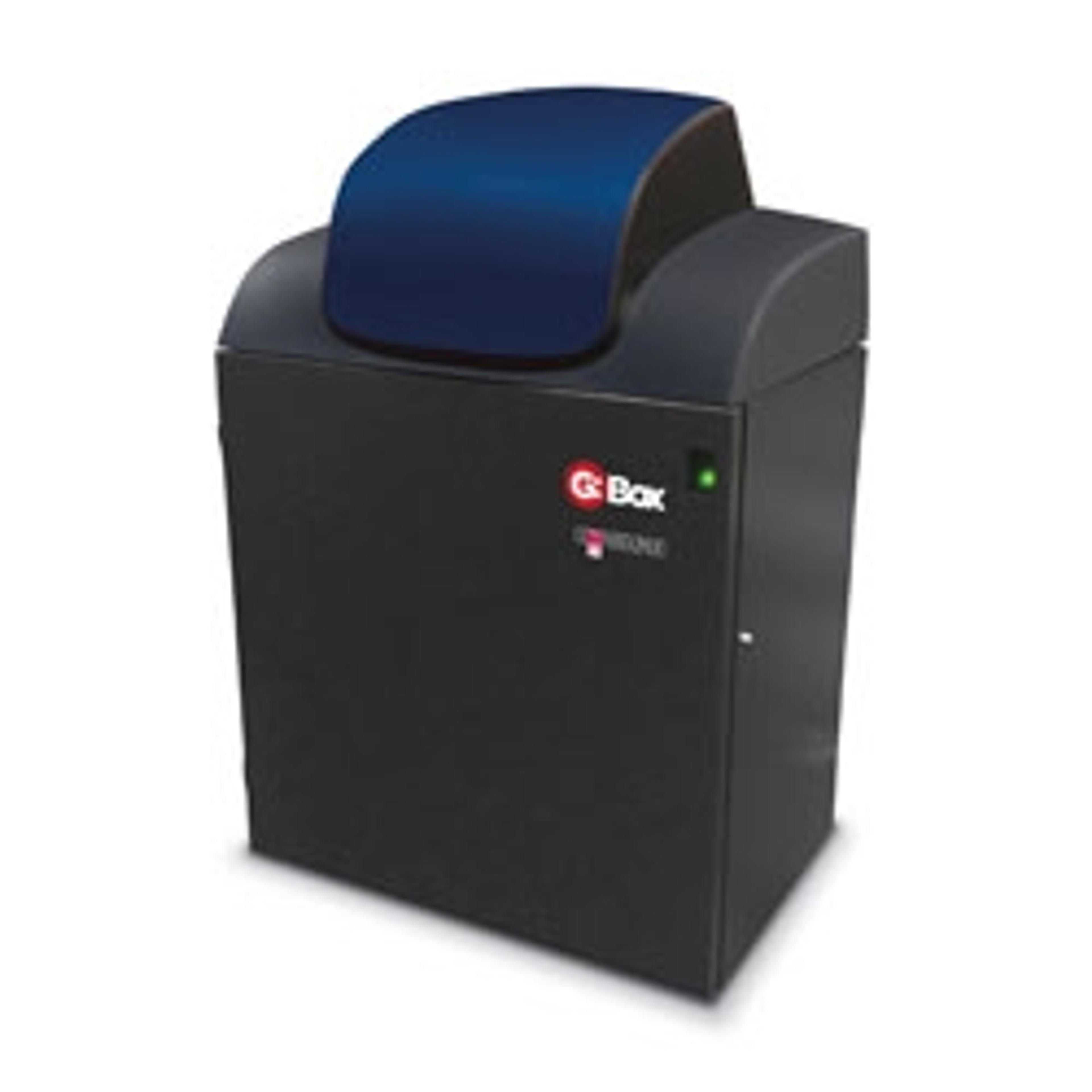
Syngene's G:BOX XR5 is Being Utilized at The University of Cambridge
10 Sept 2012Syngene, a world-leading manufacturer of image analysis solutions, is delighted to announce that the G:BOX XR5 imaging system is now being used at the University of Cambridge. The G:BOX XR5 imaging system is being used to visualize and analyze DNA as part of a research program to understand the molecular mechanisms behind why the remyelination process fails in diseases such as Multiple Sclerosis (MS).
Scientists in the Neurosciences Group at the Department of Veterinary Medicine, part of the University of Cambridge are using a G:BOX XR5 system to accurately image gels of fluorescent dye stained PCR products derived from genes involved in the remyelination process. By studying these genes, the scientists hope to find a means of enhancing the repair of normally non-repairable clinical conditions such as MS.
Dr Chao Zhao Assistant Director of Research, in the Neurosciences Group says: “Our research focuses on the mechanism of remyelination of the central nervous system after demyelination. To do this we are trying to understand the molecular mechanism of why myelin repair fails in diseases such as MS and we use the G:BOX XR5 every day to visualize and sometimes analyze very small amounts of PCR products as part of this research.”
Dr Zhao continued: “We consistently get the results we want with our G:BOX XR5 so we are happy with the system for our DNA analysis work.”
Laura Sullivan, Syngene’s Divisional Manager said: “We are delighted to hear researchers at such a prestigious department in the university are using Syngene technology in this important program. Their work is testament to the fact that for DNA analysis, the G:BOX XR5 is a totally reliable and sensitive gel documentation system.”

