The breakthrough technology transforming RNA detection
Dr. Boye Schnack Nielsen shares tips for running successful in situ hybridization assays as he provides insights on how RNA detection methods have evolved over time
25 May 2021

Diagnostic tools in histopathology have traditionally focused on studying DNA and chromosome abnormalities and measuring protein levels. Polymerase chain reaction (PCR) and microarrays, that leverage the stability of DNA and protein molecules respectively, have become established gold standards to study molecular profiles in various diseases. However, molecular scientists have long been aware that they are losing out on clinically relevant information when they are skipping the rich context that RNA analysis provides in variations of gene expression patterns.
Filling the gaps
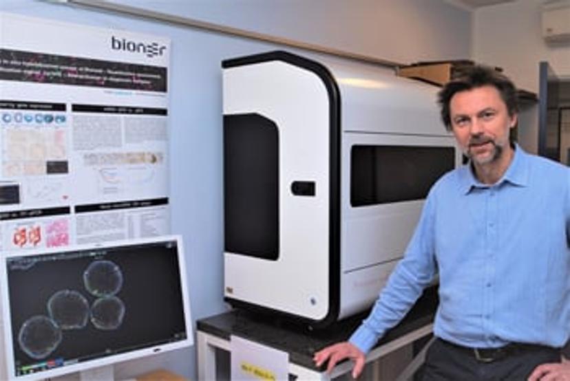
Helping to address this information gap is a key focus for molecular histologists such as Dr. Boye Schnack Nielsen, who is a Business and Research Manager for Molecular Histology at Bioneer A/S. “RNAs are interesting groups of molecules including the transfer RNAs (tRNA) and ribosomal RNAs (rRNA), that are more well known, along with the long non-coding RNAs (lncRNA), microRNAs (miRNA), and circular RNAs (circRNA), that have more recently risen to prominence. Conventionally, RNAs have not been considered as diagnostic tools because RNAs are easily lost due to degradation,” says Nielsen. “Moreover, RNA detection methods have often been complicated, necessitating the presence of skilled lab technicians.”
At Bioneer, a technological service provider approved by the Danish Ministry of Science and Technology, Nielsen and his team help companies surpass such challenges. Ongoing projects include developing and optimizing RNA detection methods such as RNA in situ hybridization assays and combining them with immunohistochemistry techniques. As a contract research organization, Bioneer works alongside its biotechnology clients to execute full lifecycle translational projects in disease biology. Such projects typically involve addressing questions in oncology, CNS and stem cell research, as well as developing and producing drug candidates for preclinical studies.
The secret to success: Control assays
“Key to successful RNA work is to always run control assays,” says Nielsen. “In case of a failed experiment, it can be tricky to narrow down the culprit amongst incorrectly processed samples, inadequate experimental conditions, and RNA probes with poor detection quality. Even the two types of tissues used for RNA work, frozen and paraffin-embedded tissues, can both give good results provided they are processed properly,” he continues. “When your assay is not working as expected, you need to test different experimental conditions out. For this, you rely on your panel of control assays to help resolve the issue. I use a combination of in-house positive control tissue to test my sample quality and control probes to test the performance of my probes.”
Over the past decade, the field of RNA diagnostics has evolved rapidly, expanding the scope for applying RNA detection techniques, Nielsen asserts. “Companies like Advanced Cell Diagnostics (ACD) have developed innovate methods of RNA detection for use in histology,” explains Nielsen. “To ensure ease of implementation, these RNA assays are adapted for automated use on Leica and [Roche] Ventana instruments which are commonly seen in pathology labs. Focusing on delivering sensitivity and specificity, these assays have also become more reproducible over the years.”
One of the prominently used methods for miRNA staining in clinical samples has been LNA probe technology since its introduction in 20101. Making use of nucleic acid analogs called locked nucleic acids, LNA technology uses both chromogenic and fluorescent methods to detect a range of RNA as well as DNA molecules. Whilst LNA has significantly higher sensitivity and target specificity compared to DNA and RNA probes used in the past, reproducibility can still be tricky for some of the shorter RNA molecules.
An optimized, reliable assay
Aiming to tackle this shortcoming of LNA technology, assay developers at ACD designed the miRNAscope™, an in situ hybridization assay. The miRNAscope helps visualize antisense oligonucleotides (ASOs), miRNAs, small interfering RNAs (siRNAs), and other short nucleic acid targets in both intact tissues as well as cultured cells.
As one of the first pioneers of the LNA technology, Nielsen knew that for the miRNAscope to be a true improvement on LNA technology, to begin with, it had to replicate the latter’s results. To assess the miRNAscope, his team used targets that typically get picked up well via LNA such as U6, a small nuclear RNA, and microRNA-126, an endothelial-specific microRNA. And the miRNAscope passed the test with flying colors, explains Nielsen: “We know what to look for in the staining patterns: with the miRNAscope, we see a clear localization of microRNA-126 in the blood vessels. Moreover, for another microRNA that was not detected in a reproducible way with the LNA technology, we see better output with the miRNAscope. This is not entirely unexpected considering the tremendous optimization efforts that have gone into making the miRNAscope more sensitive,” he says.
Future outlooks
Nielsen is excited that the miRNAscope empowers companies such as Bioneer as well as their clients with an alternative for detecting microRNAs, other than just via LNA. Moving forward, he foresees that miRNAscope will be a significant improvement on LNA assays, enabling better staining of a wider range of microRNAs and improving assay reproducibility. “We highly appreciate the innovative staff in companies like ACD, who are not only focused on designing probes and improving detection technology but are also keen on refining novel tissue processing steps,” says Nielsen. As he looks forward to using the automated version of the miRNAscope kit next, Nielsen concludes that he is hopeful that the full potential of RNA detection in the field of disease biology will soon be realized.
References
1. Jørgensen, S., Baker, A., Møller, S., & Nielsen, B. S. Robust one-day in situ hybridization protocol for detection of microRNAs in paraffin samples using LNA probes. Methods, 2010.
Do you use Advanced Cell Diagnostics products in your lab? Write a review for your chance to win a $400 Amazon gift card>>

