Uncover the cutting-edge technologies in the field of microscopy
Explore AI-powered image analysis, Raman microscopy and energy-dispersive X-ray spectrometry and how they can be utilized across a range of scientific applications
7 Aug 2023Microscopy enables scientists to look beyond what is visible to the human eye, unravelling the hidden characteristics of living and non-living things. What started with a simple light microscope, has advanced beyond imagining in recent years. Now scientists have technology choices including 3D electron microscopes, scanning transmission electron microscopy, corelative light and electron microscopy, and electron backscatter diffraction (EBSD).
AI within the microscopy field: A second pair of eyes
Artificial intelligence has the potential to revolutionize microscopy image analysis. By training ‘smart microscopy’ tools to achieve reliable object classification, image classification, segmentation, image restoration, super resolution, and virtual staining, scientists working in fields such as patient diagnostics and cancer research are benefitting from higher resolution and more immediate insights.
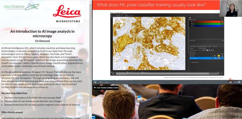
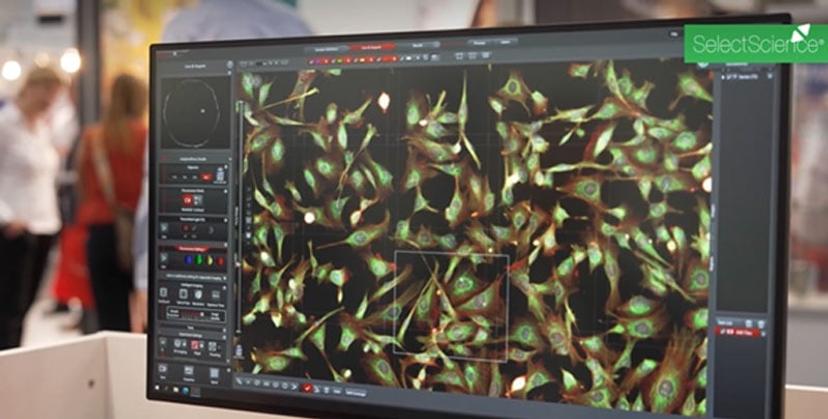
An introduction to AI image analysis in microscopy
Over the past four years, there has been a trend towards microscopists using AI- based solutions for image acquisition optimization (smart microscopy), object classification, image classification, segmentation, restoration, super resolution, and virtual staining.
In this educational webinar, AI expert, Dr. Quyen Tran, Leica Microsystems, discusses the basic concepts and terminology of AI, defining some of the key terminology that can be hard to decipher for non-AI experts. Sharing how machine and deep learning can be used for microscopy applications, Dr. Tran demonstrates how we can extract further insights from our image acquisition and analysis.
AI-based image analysis algorithms
Medical imaging generates vast amounts of data that are impossible to analyze manually. With advances in AI, researchers are now exploring how this technology can be used to manage and simplify the analysis of large 3D microscopy data sets.
In this article, Jianxu Chen, head of the new Analysis of Microscopic BIOMedical Images (AMBIOM*) group at ISAS discusses how his research group is developing scalable, AI-based image analysis algorithms to support disease studies.
Discover Chen’s research goals, find out how machine learning and AI are helping to overcome technological challenges, and find out the potential impact his team’s work will have in the field of healthcare.
Analytical microscopy: Full characterization of samples
Full characterization of organic and inorganic samples provides a better understanding of their properties and structure. Combining different microscopic techniques and pushing the boundaries of current technologies can achieve full characterization which helps research and applications in different sectors including pharmaceutical, environmental, and industrial.
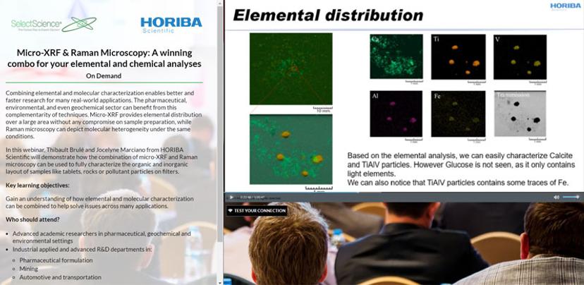
Micro-XRF & Raman microscopy: A winning combo
In this webinar Thibault Brulé and Jocelyne Marciano from HORIBA Scientific demonstrate how the combination of micro-XRF and Raman microscopy can be used to fully characterize the organic and inorganic layout of samples like tablets, rocks, or pollutant particles on filters.
Understand how elemental and molecular characterization can be combined to help solve issues across many applications.
Electron backscatter diffraction: Moving toward the mainstream
In this article, Dr. Pat Trimby, EBSD Product Manager at Oxford Instruments discusses the advances in electron backscatter diffraction (EBSD) that will make it more accessible for researchers in different fields such as biomaterials and green energy. The article explains how the use of high-speed complementary metal oxide semiconductor (CMOS) technology in EBSD detectors instead of the standard charge-coupled device (CCD) sensors, can produce higher resolution images at a high speed.
Discover how the combination of EBSD with energy-dispersive X-ray spectrometry (EDS), provides comprehensive analysis of a sample’s physical properties and how this can be applied across different industries and research areas, from metals research to advanced manufacturing techniques, renewable energy, microelectronics, and geological research.
A dedicated EBSD learning center is available from Oxford Instruments for researchers that want to enhance their understanding of the advancements of the technology and its future applications.
Super-resolution imaging: Unfold the secrets of biology
Explore the power of super-resolution microscopy: Unveiling the unseen
In this interview, with Winfried Wiegraebe, Ph.D., Product Manager of Super-Resolution Microscopy at Bruker, discover the benefits of this super-resolution microscopy, understanding how it works and its applications in different research fields, as well as learning about one of Bruker’s super-resolution microscopes.
Wiegraebe discusses single-molecule localization microscopy (SMLM) and its ability to collect data from individual molecules within a sample, overcoming the diffraction limit of conventional microscopes producing super resolution images with the most intricate details.
Step into the future of microscopy
The future of microscopy: Latest innovations from Leica Microsystems
Watch this video to learn about the key features and research applications of the microscopy product portfolio available from Leica Microsystems – from the ‘world’s first imaging Microhub’ to cryo-immobilization of aqueous samples.
Watch the full video interview.
Find more news and resources in our Accelerating Science Special Feature>>