AMH (Anti-Mullerion Hormone) ELISA
High Quality Assays with Reproducible and Reliable Results

The supplier does not provide quotations for this product through SelectScience. You can search for similar products in our Product Directory.
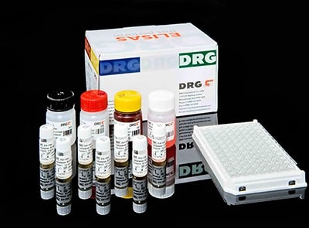
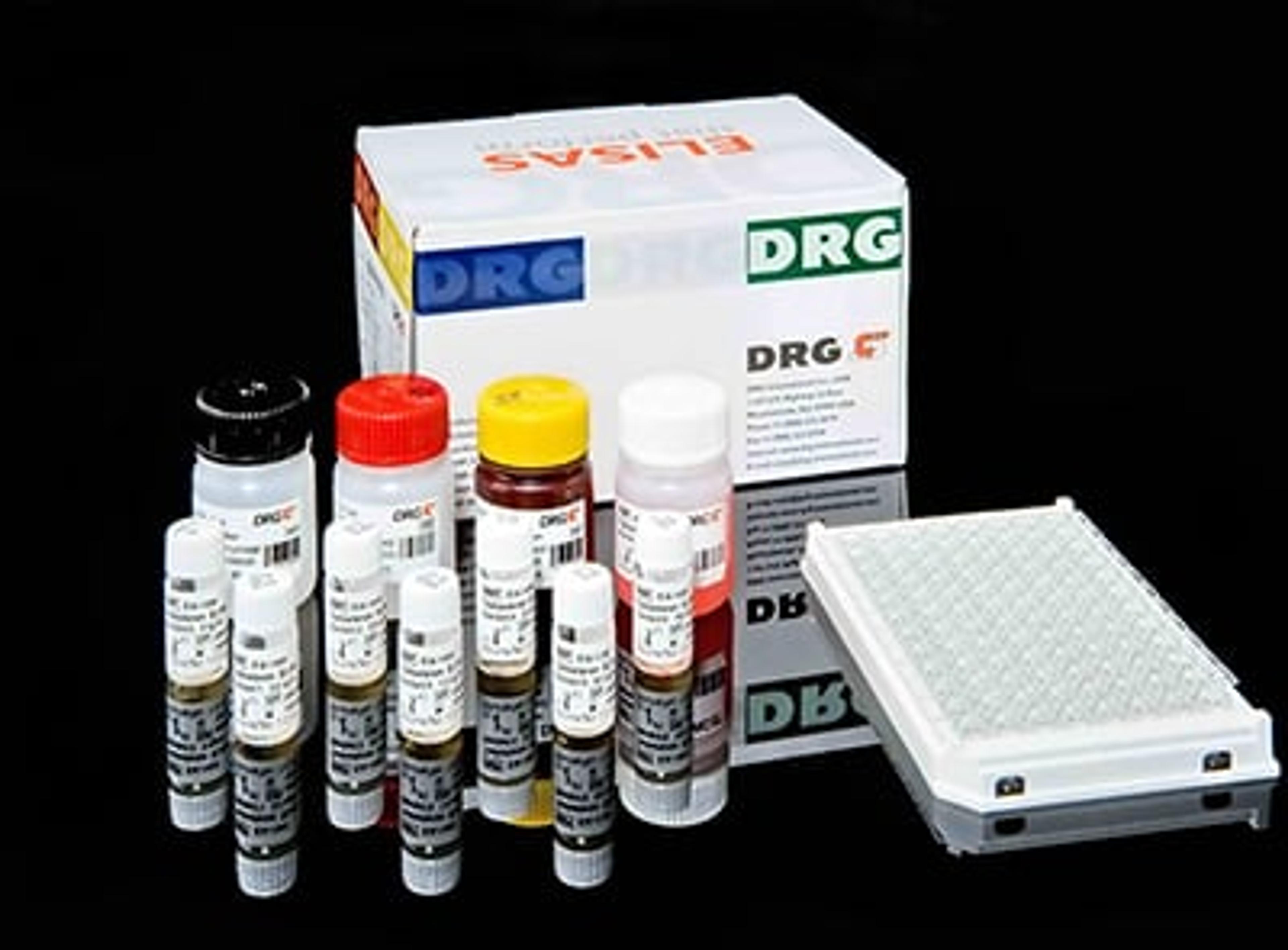
An enzyme immunoassay for the quantitative measurement of Anti-Müllerian Hormone in serum and plasma. Anti-Müllerian hormone also known as AMH is a protein that, in humans, is encoded by the AMH gene. (1) It inhibits the development of the Müllerian ducts (paramesonephricducts) in the male embryo (2). It has also been called Müllerian inhibiting factor (MIF), Müllerian-inhibiting hormone (MIH), and Müllerian-inhibiting substance (MIS). AMH is a protein hormone structurally related to inhibin and activin, and a member of the transforming growth factor-Beta (TGF-Beta) family. It is a dimeric glycoprotein that has a molar mass of 140 kDa (3). AMH is secreted by Sertoli cells of the testes during embryogenesis of the fetal male. In females, it is secreted by the granulosa cells of ovarian follicles. In mammals, AMH prevents the development of the müllerian ducts into the uterus and other müllerian structures (2). The effect is ipsilateral, that is each testis suppresses Müllerian development only on its own side. (4) In humans, this action takes place during the first 8 weeks of gestation. AMH is expressed by granulosa cells of the ovary during the reproductive years, and controls the formation of primary follicles by inhibiting excessive follicular recruitment by FSH. It, therefore, has a role in folliculogenesis (5), and some authorities suggest it is a measure of certain aspects of ovarian function, (6) useful in assessing conditions such as polycystic ovary syndrome and premature ovarian failure (7). It is useful to predict a poor ovarian response in in vitro fertilization (IVF), but it does not appear to add any predictive information about success rates of an already established pregnancy after IVF (8). In men, inadequate embryonal AMH activity can lead to the Persistent Müllerian duct syndrome (PMDS), in which a rudimentary uterus is present and testes are usually undescended. The AMH gene or the gene for its receptor are usually abnormal. AMH measurements have also become widely used in the evaluation of testicular presence and function in infants with intersex conditions, ambiguous genitalia, and cryptorchidism. Its ability to inhibit growth of tissue derived from the Müllerian ducts has raised hopes of usefulness in the treatment of a variety of medical conditions including endometriosis, adenomyosis, and uterine cancer.The DRG AMH ELISA is a quantitative solid phase enzyme-linked immunosorbent assay (ELISA), based on the principle of a sandwich type immunoassay. The microtiter wells are coated with an AMH antibody. Standards, Controls and unknown samples are added to the microtiter wells and incubated. The wells are then washed, and biotinylated AMH antibody solution is added and the wells are incubated. The antibody-biotin conjugate binds to the solid phase antibody - antigen complex. After incubation and washing, streptavidin horseradish peroxidase conjugate (SHRP) solution is added to the wells and incubated while the biotin binds to the streptavidin-enzyme conjugate. The wells are again washed and tetramethylbenzidine (TMB) substrate solution is added and incubated, followed by an acidic stop solution. The antibody-antigen-biotin conjugate-SHRP complex bound to the well is detected by enzyme-substrate reaction. The intensity of color developed is proportional to the concentration of AMH in the patient sample measured at 450 nm as primary test filter and 630 nm as reference filter. Values obtained from the absorbance readings are calculated from a Standard Curve generated using the absorbance of the Standards.

