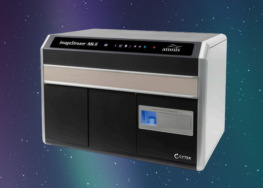
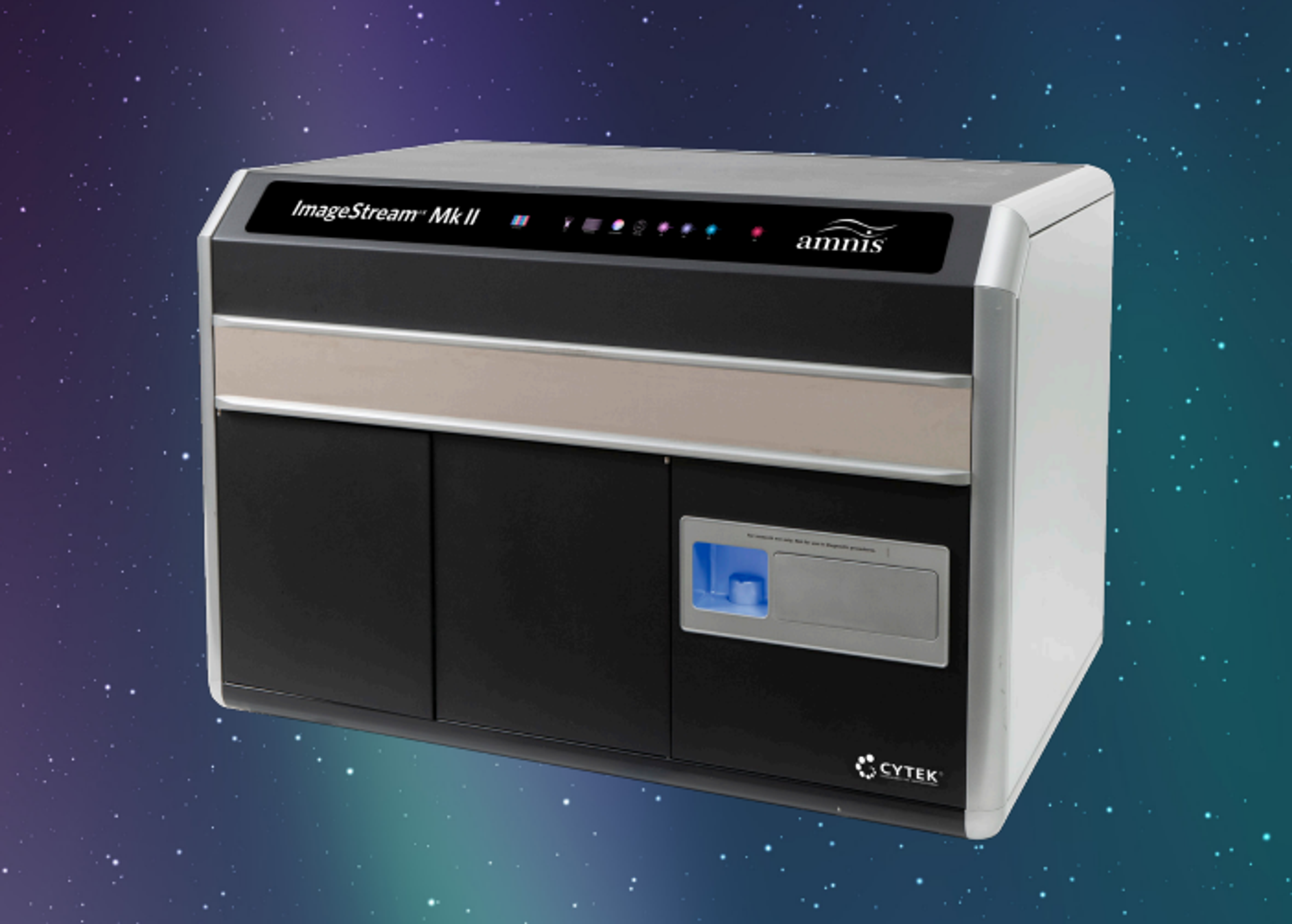
Amnis® ImageStream® XMk II
The revolutionary ImageStream ® X Mk II Imaging Flow Cytometer combines the speed, sensitivity, and phenotyping abilities of flow cytometry with the detailed imagery and functional insights of microscopy. This unique combination enables a broad range of applications that would be impossible using either technique alone.

The supplier does not provide quotations for this product through SelectScience. You can search for similar products in our Product Directory.
Excellent data quality, unique applications
Imaging Flow Cytometry
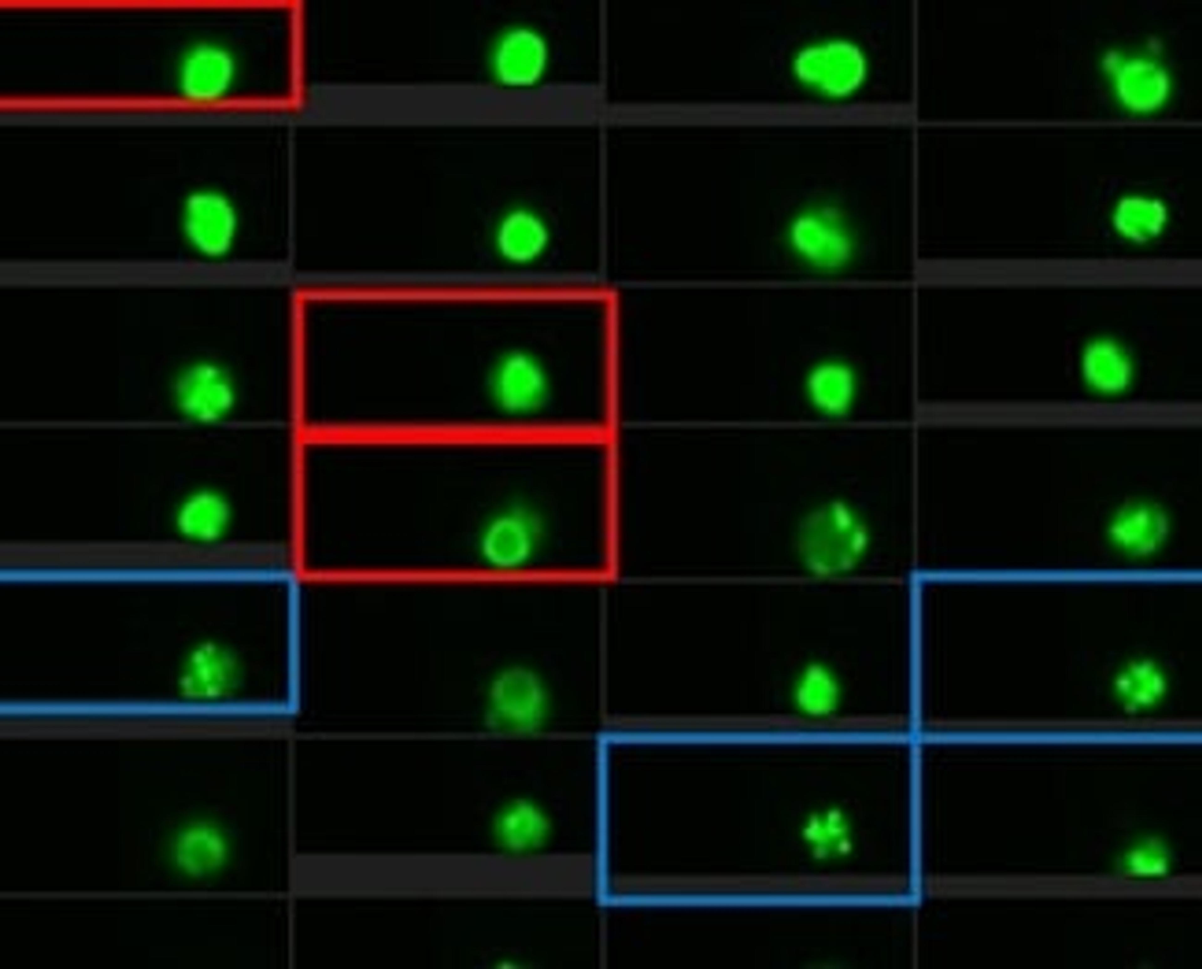
The instrument combines the information-rich imagery of microscopy with the high-throughput rapid acquisition of flow cytometry. It allows acquisition of bright-field and up 10 fluorescent images of cells in flow, up to 5000 cells / second. It has 20X, 40X and 60X magnification, the minimal pixel size is 330nm. Among the common application are co-localization, internalization, morphology, spot count and many others. The data analysis is done by a dedicated image analysis software, IDEAS. It allows robust, un-biased high throughput quantification of morphological features. I have been using it for the last 12 years, resulting in over 100 publications in numerous disciplines in biology, including immunology, cancer, cell biology, microbiology, marine biology, and many others. I can highly recommend it for all cellular applications.
Review Date: 20 Oct 2023 | Cytek Biosciences
Easy to use and versatile
Cellular biomarkers
Easy to use. It does not require much maintenance. Good resolution and versatility. However, services are difficult to obtain due to changes in service providers.
Review Date: 20 May 2022 | Cytek Biosciences
Excellent technology for high-throughput image analysis at medium resolution.
CAR T-cell synapse analysis
This is a high-throughput image analysis platform for rare events. CAR-T-cell/target cell synapses are identified with high precision and efficiency. The company provide extensive support for application advice and troubleshooting. Probably the highest-throughput image analysis system for its price.
Review Date: 29 May 2020 | Cytek Biosciences
The Cytek® Amnis® ImageStream® XMk II imaging flow cytometer combines the high-throughput performance of flow cytometry with the imagery and functional insights of microscopy. This unique combination enables a broad range of applications that would be impossible using either technique alone.
The ImageStream XMk II system is a benchtop, multispectral, imaging flow cytometer designed for the rapid acquisition of millions of cells with up to 12 channels of cellular imagery. The instrument collects large numbers of digital images, performs spectral compensation, and provides high-quality images of every cell in your sample. Combined with our advanced image analysis software, where each object is measured for hundreds of parameters using not only fluorescence intensity but the fluorescence spatial location as well, the ImageStream system offers an unprecedented level of cellular information.

