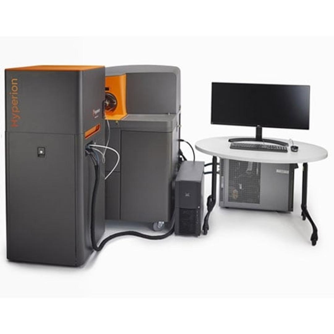
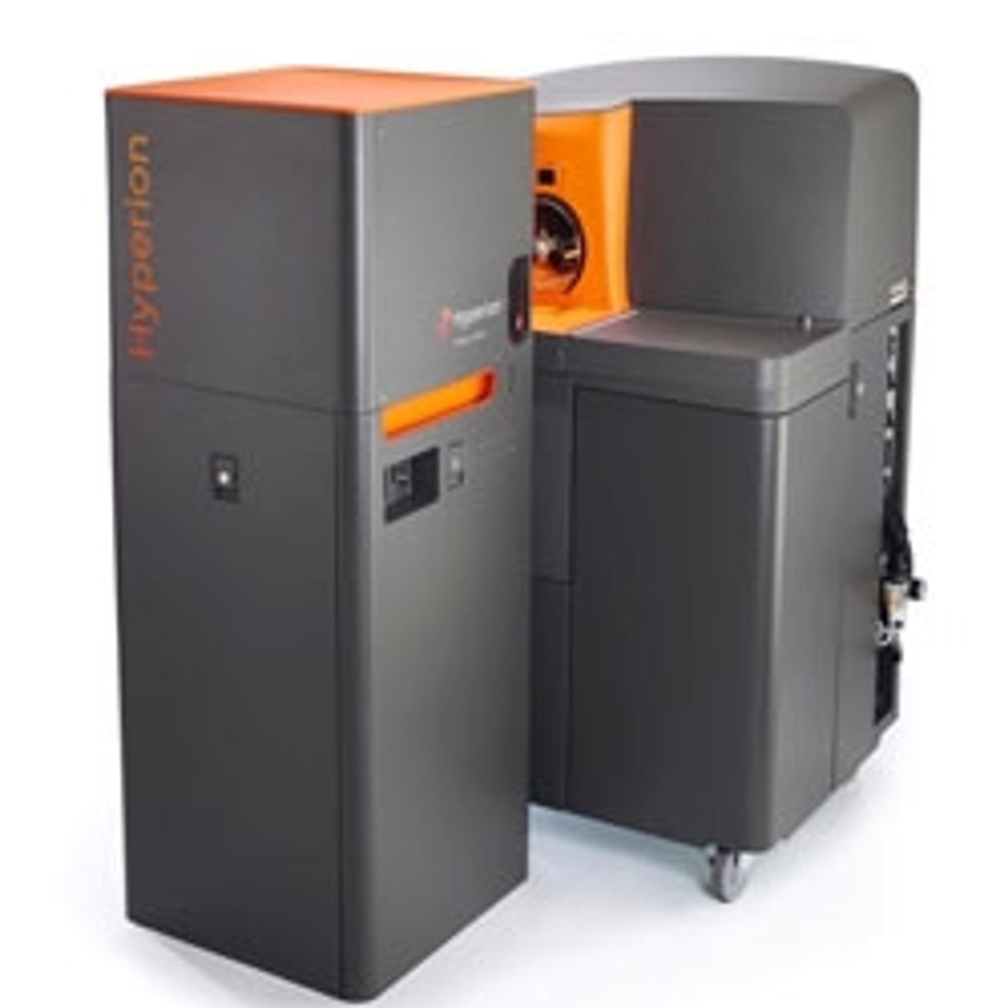
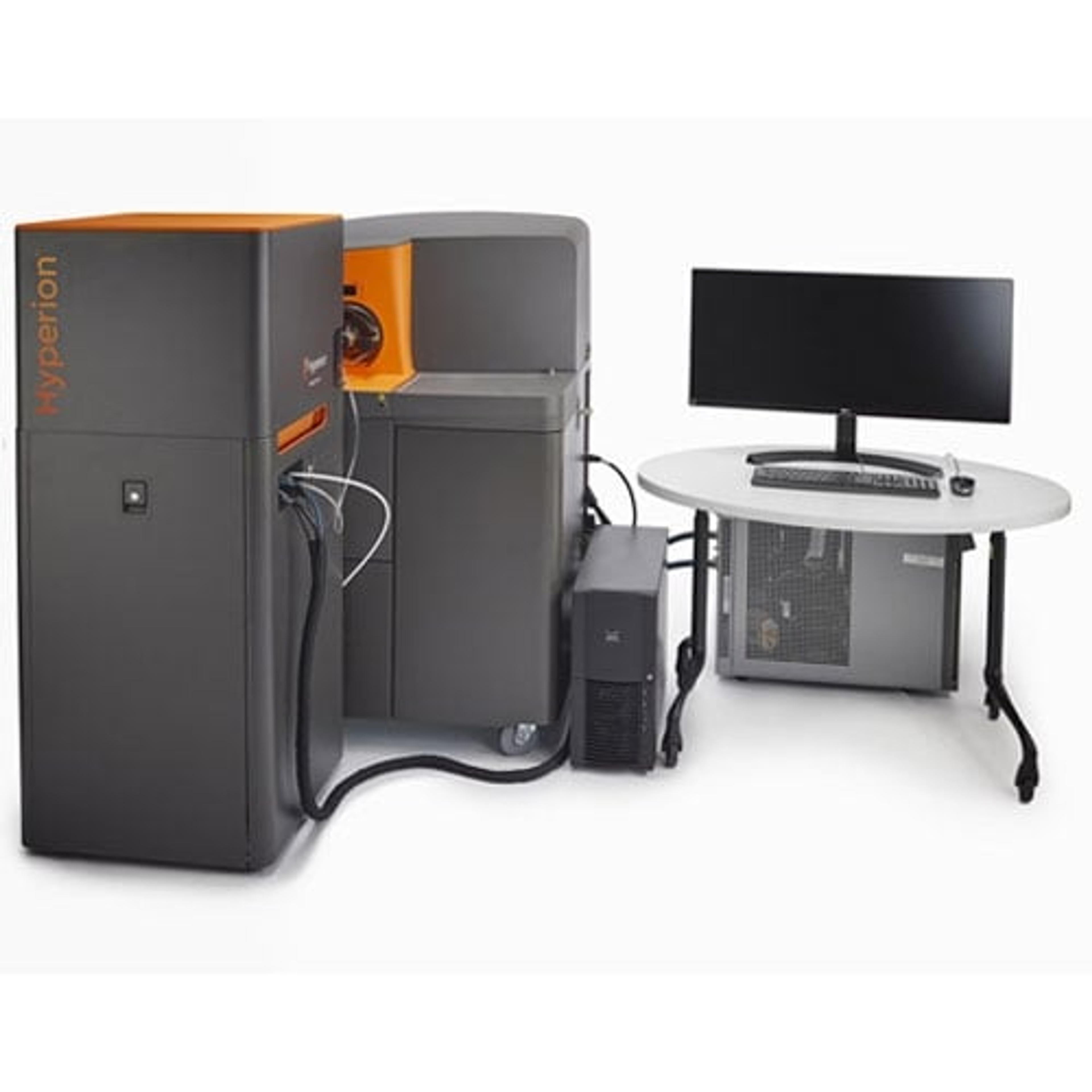
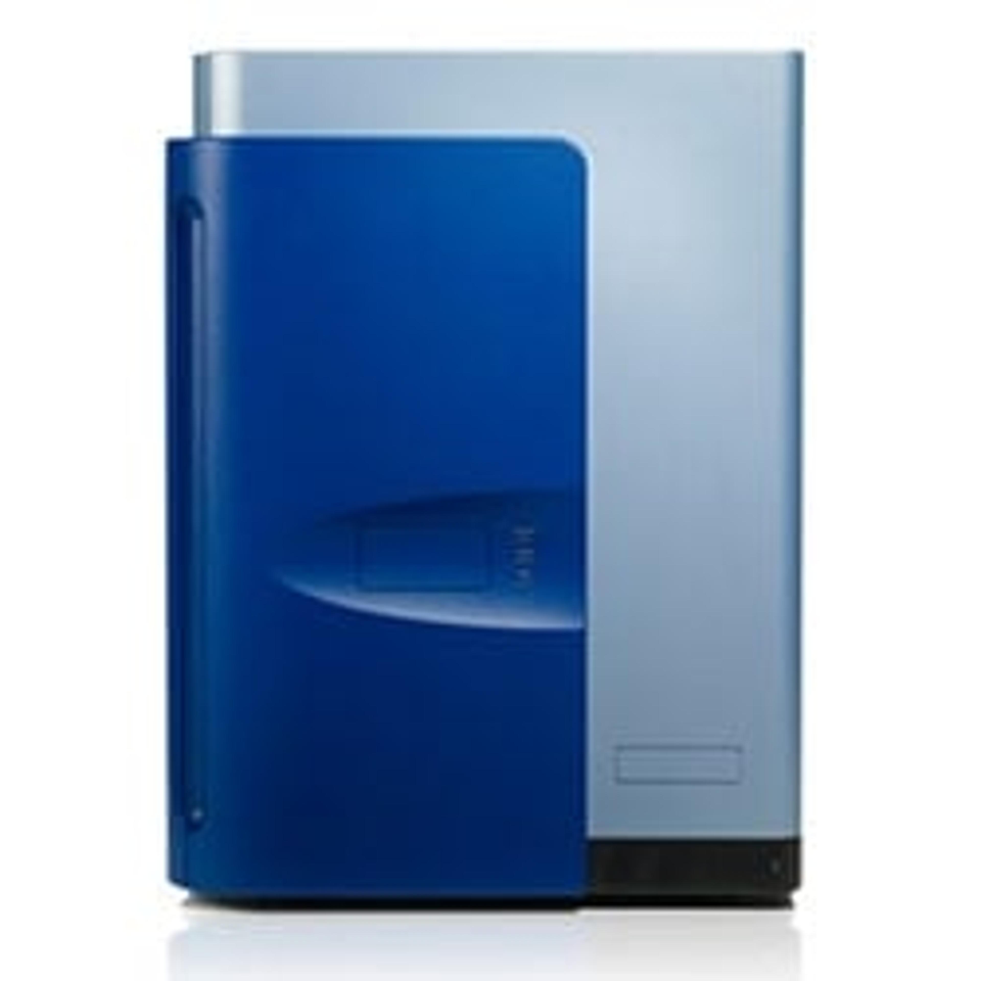
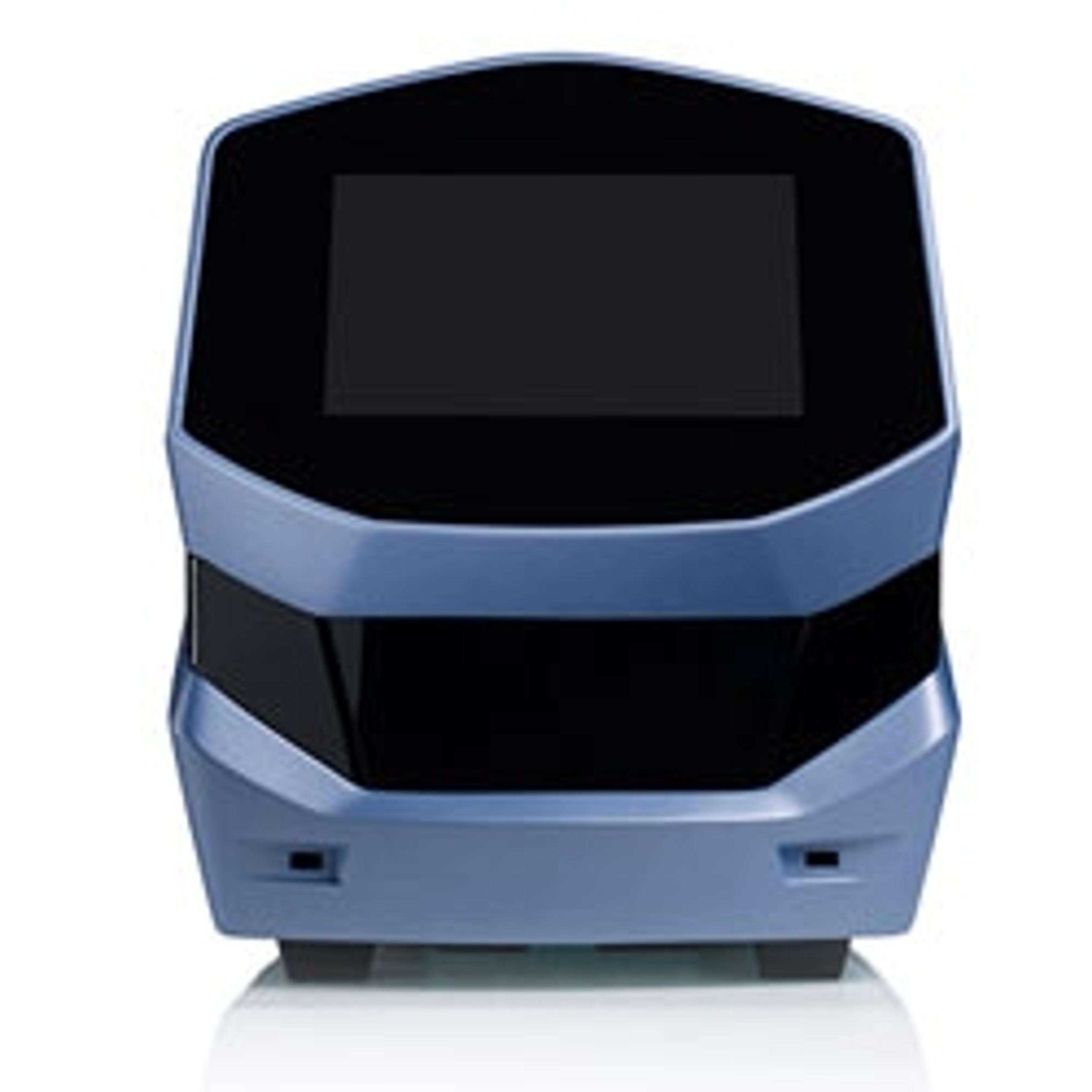
Hyperion™ Imaging System
A transformative imaging solution that enables comprehensive analysis of cellular phenotypes and their interrelationships.
Great results, but need time for analyses
Analyze tumor microenvironment
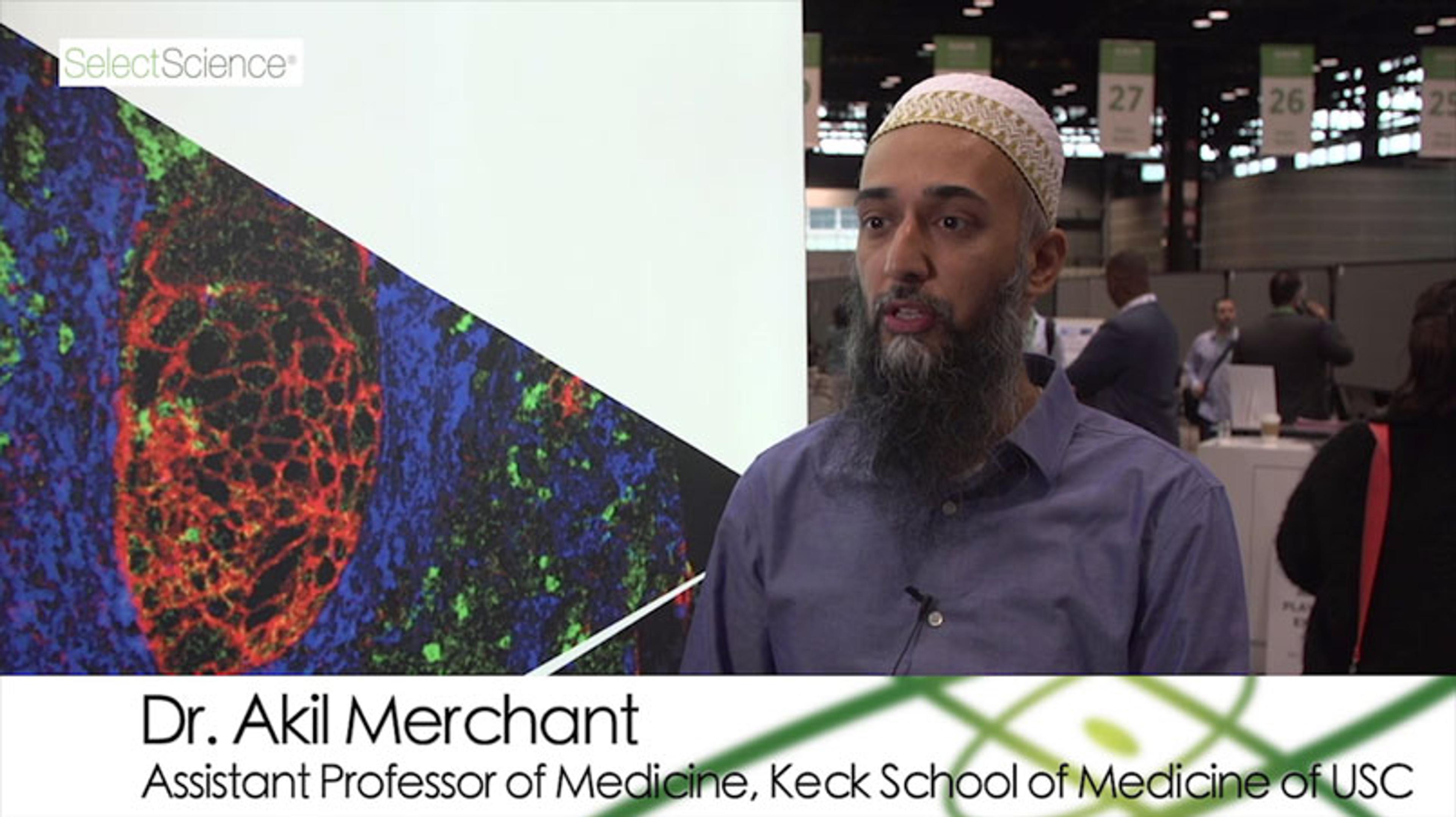
The automat has been used for the exploration of the tumoral microenvironment of tumoral and inflammatory colon and gastric tissues. Acquisitions take a bit of time, but it is quite easy to use the automat. The longest part is to make the preparation work for the antibodies panel and the tissues, and the most difficult part is the analysis of the huge amount of data generated by Hyperion.
Review Date: 8 Jun 2022 | Standard BioTools Inc.
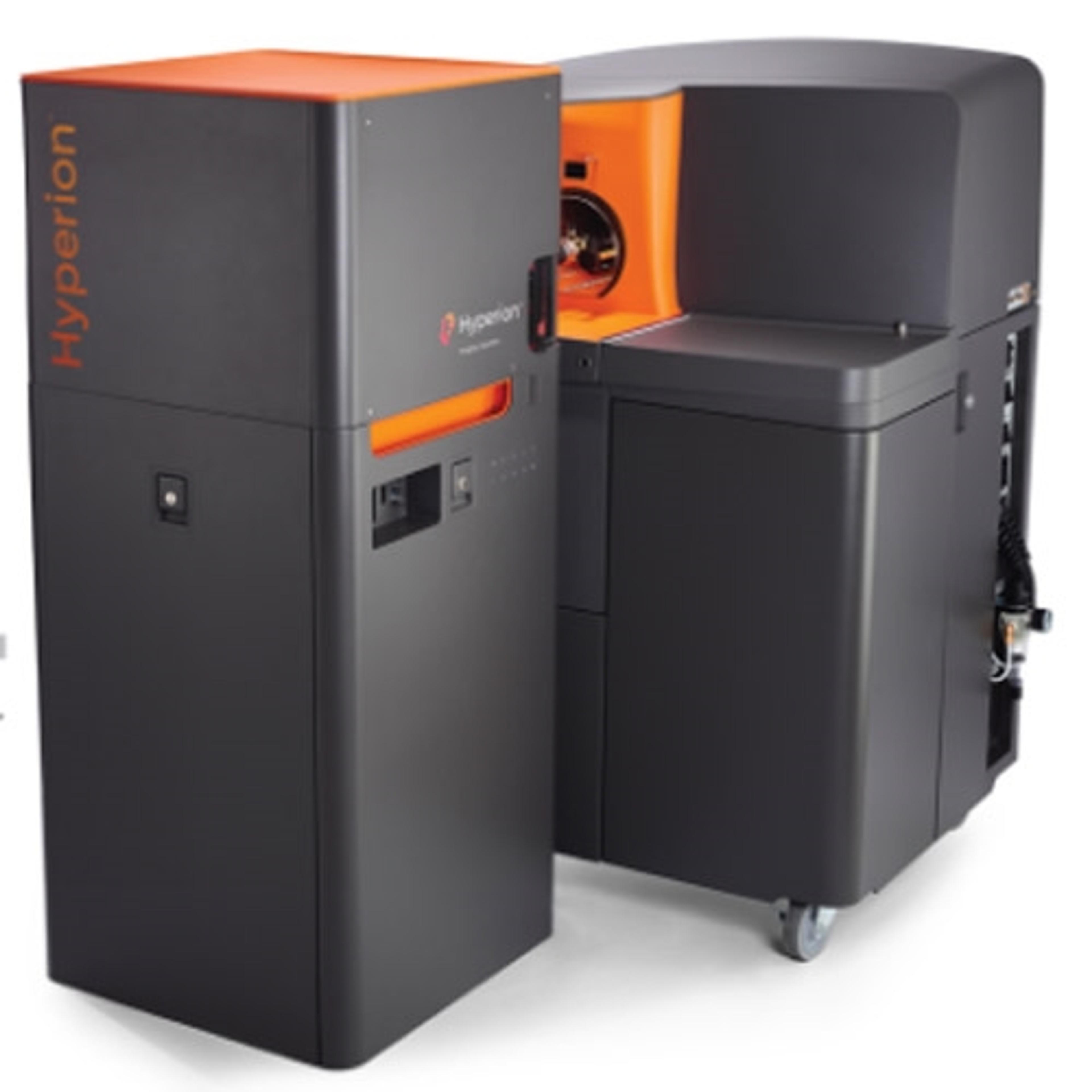
Changing the course of how diseases are treated and ultimately cured requires a comprehensive understanding of complex cellular phenotypes and their interrelationships in the spatial context of the tissue microenvironment. Imaging Mass Cytometry™ (IMC™) performed on the Hyperion™ Imaging System using Maxpar® metal-tagged antibodies empowers simultaneous imaging of up to 37 protein markers imaged at a time. Bringing together high-parameter CyTOF® technology with imaging capability, the Hyperion Imaging System enables deep interrogation of tissues and tumors at subcellular resolution to uncover pathology insights, new biomarker correlations and cell interactions.
- Comprehensive - Highly multiplexed immunohistochemistry (IHC) using proven CyTOF technology enables simultaneous analysis of 4 to 37 protein markers from a single scan
- Contextual - Identify new protein biomarkers and deeply profile tissue microenvironments at subcellular resolution while preserving the information in tissue architecture and cellular morphology
- Powerful - Preserve precious samples and reduce variability by eliminating dependency on serial sections

