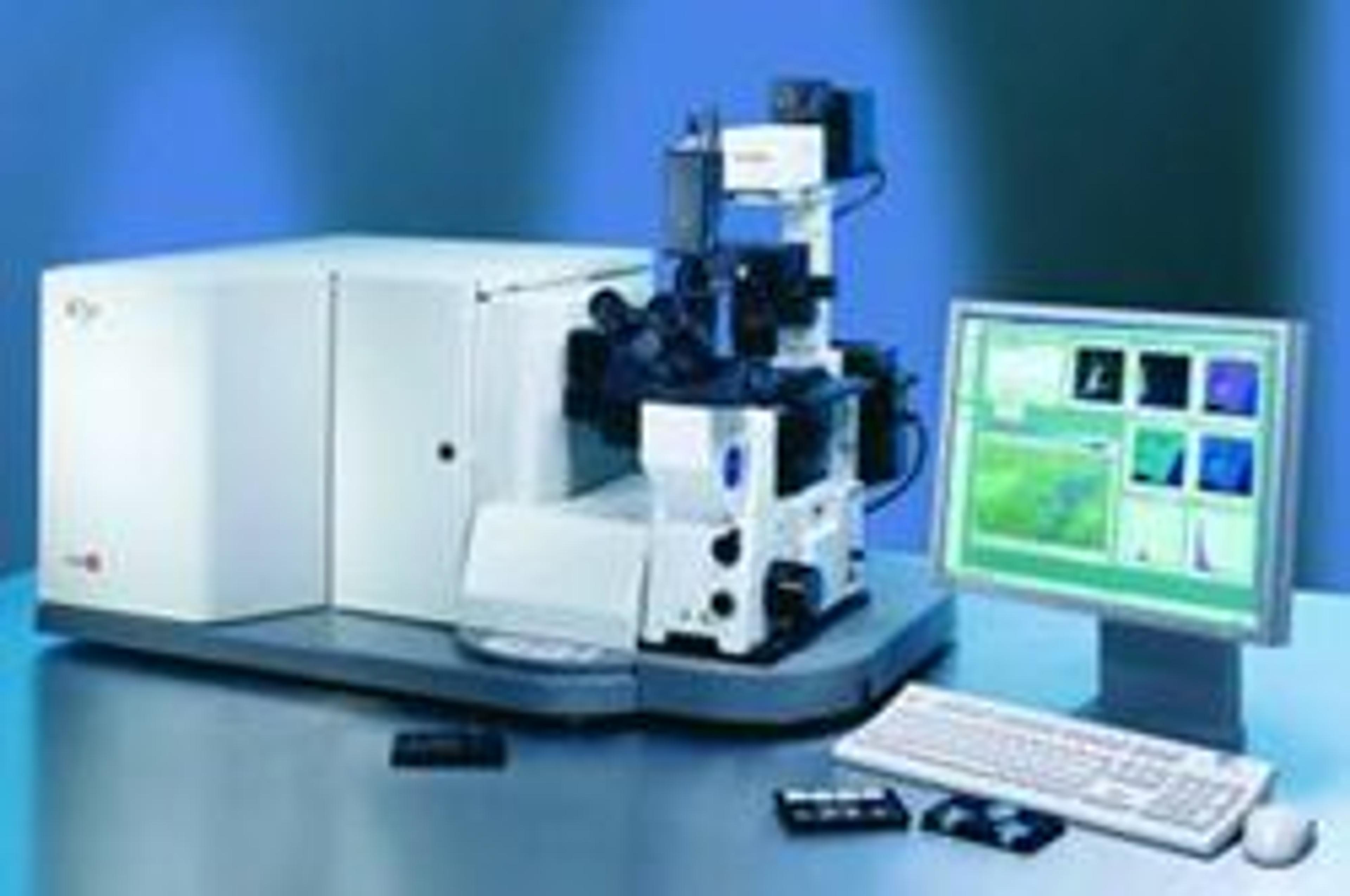
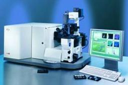
iCys® Research Imaging Cytometer
The iCys® Research Imaging Cytometer combines the technological advances of the existing LSC and iCyte instruments, providing the ability to visualize the specimen through microscope optics or with a digital camera. The combination of analytic software advances with microscope features provides the iCys with a unique mode of analysis. Once scanning and analysis are completed, the microscope stage may be automatically moved to…

The supplier does not provide quotations for this product through SelectScience. You can search for similar products in our Product Directory.
I use daily- the quality of data is good.
Review Date: 6 Jul 2011 | CompuCyte Corp.
The iCys® Research Imaging Cytometer combines the technological advances of the existing LSC and iCyte instruments, providing the ability to visualize the specimen through microscope optics or with a digital camera.
The combination of analytic software advances with microscope features provides the iCys with a unique mode of analysis. Once scanning and analysis are completed, the microscope stage may be automatically moved to the location of any event of interest. The event can then be viewed using the microscope optics while simultaneously viewing the laser scan images. The user may further utilize any number of microscope accessories available, including opto-mechanical devices for micromanipulation and cell capture.
Features include:
- Simultaneous acquisition of multiple-fluorescence and brightfield laser-scatter images, from as many as 3 lasers and 5 multiplexed light sensors
- Wide-field images, super-wide-field sampling and mosaic images
- Easily viewed specimens with on-the-fly galleries, or relocation to individual events after data acquisition
- The option for either laser scan images or live microscope or camera viewing, or both
- Analysis and visualization of specimens in a variety of formats (chamber slides, microtiter plates, petri dishes, microscope slides) to analyze cell populations, cell colonies, tissues and tissue arrays
- Precise localization of cells and tissue events of interest with the automated motorized stage
- Autofocus for automated analysis
- Easy interface with peripheral microscopy devices like cameras, phase-contrast visualization, micromanipulators, cloning devices, etc