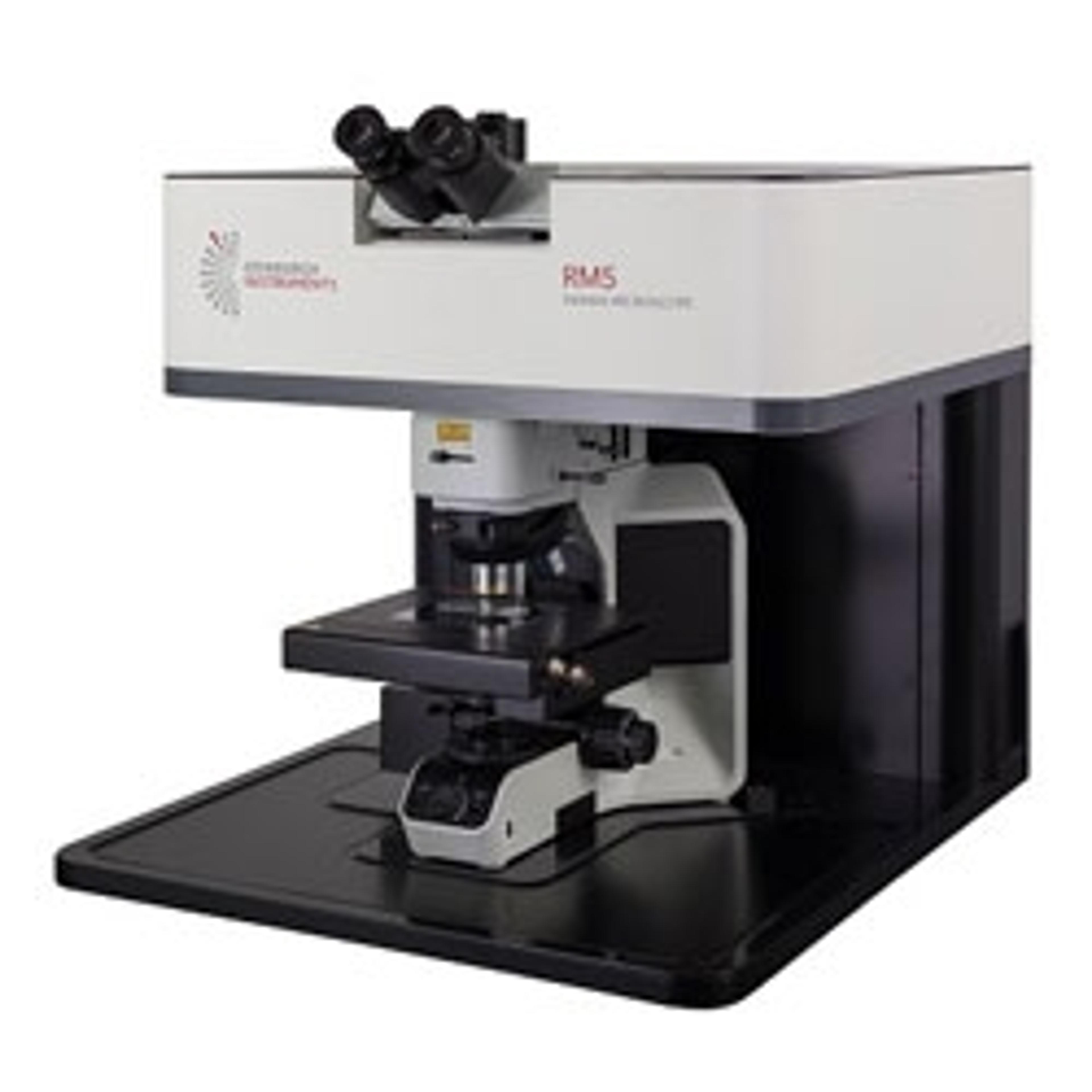
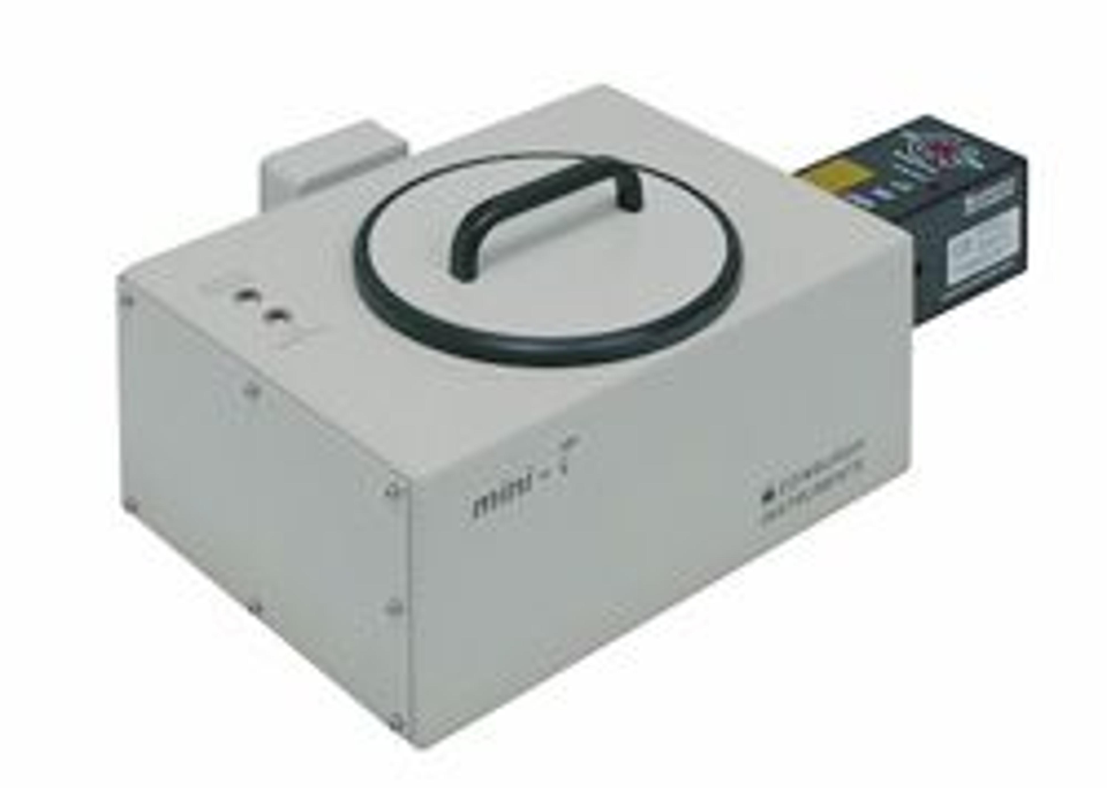
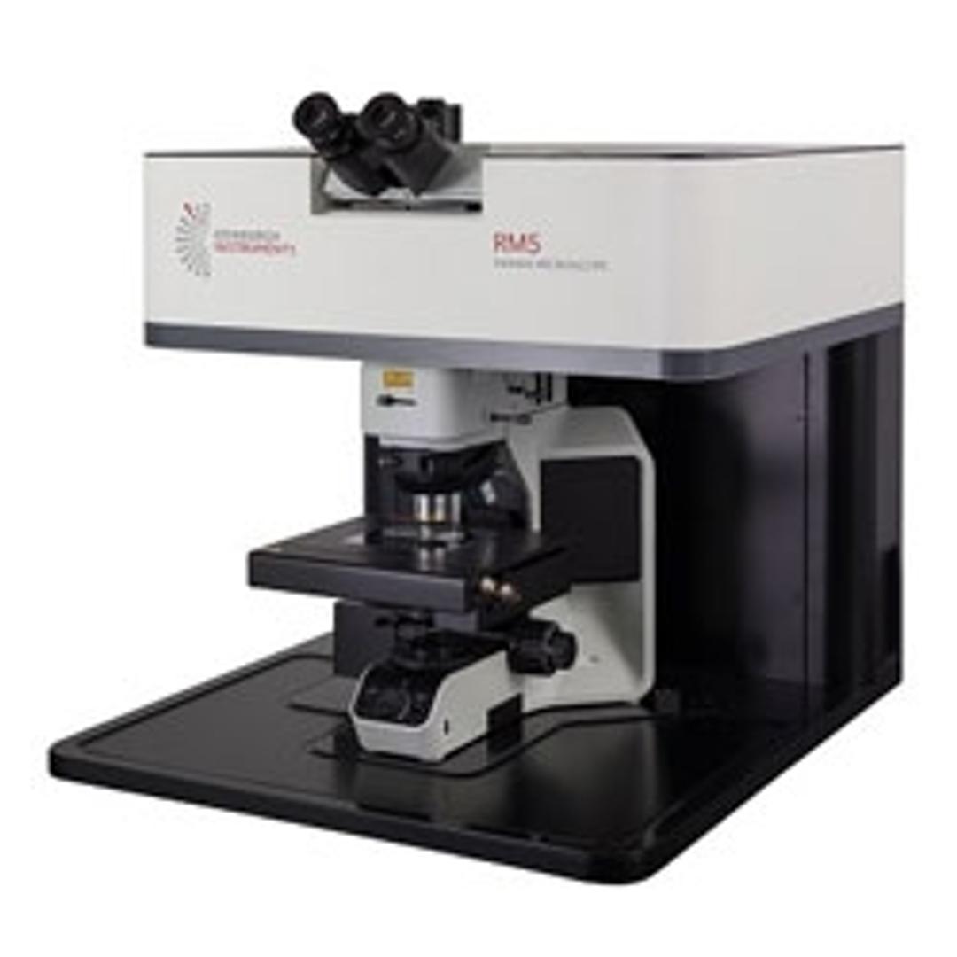
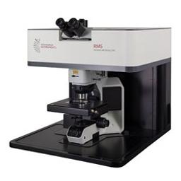
RM5 Raman Microscope
The RM5 Raman Microscope is a compact Raman microscope engineered for diverse laboratory environments, spanning industrial and academic sectors.
Spectral Measurements
2D Mapping
3D Mapping
SurfMAP®


Receive your quote directly from the manufacturer.
The RM5 Raman Microscope is a compact Raman microscope engineered for diverse laboratory environments, spanning industrial and academic sectors. Employing advanced spectral imaging, intuitive operation, and precise confocal optics, the RM5 delivers you high-performance Raman measurements across applications including materials science and pharmaceuticals.
Key Technical Features:
- Configurable Confocality: Variable slit and multi-position adjustable pinhole for enhanced image resolution, improved fluorescence rejection, and application-specific optimization. Offers a wide range of software-selectable pinhole options.
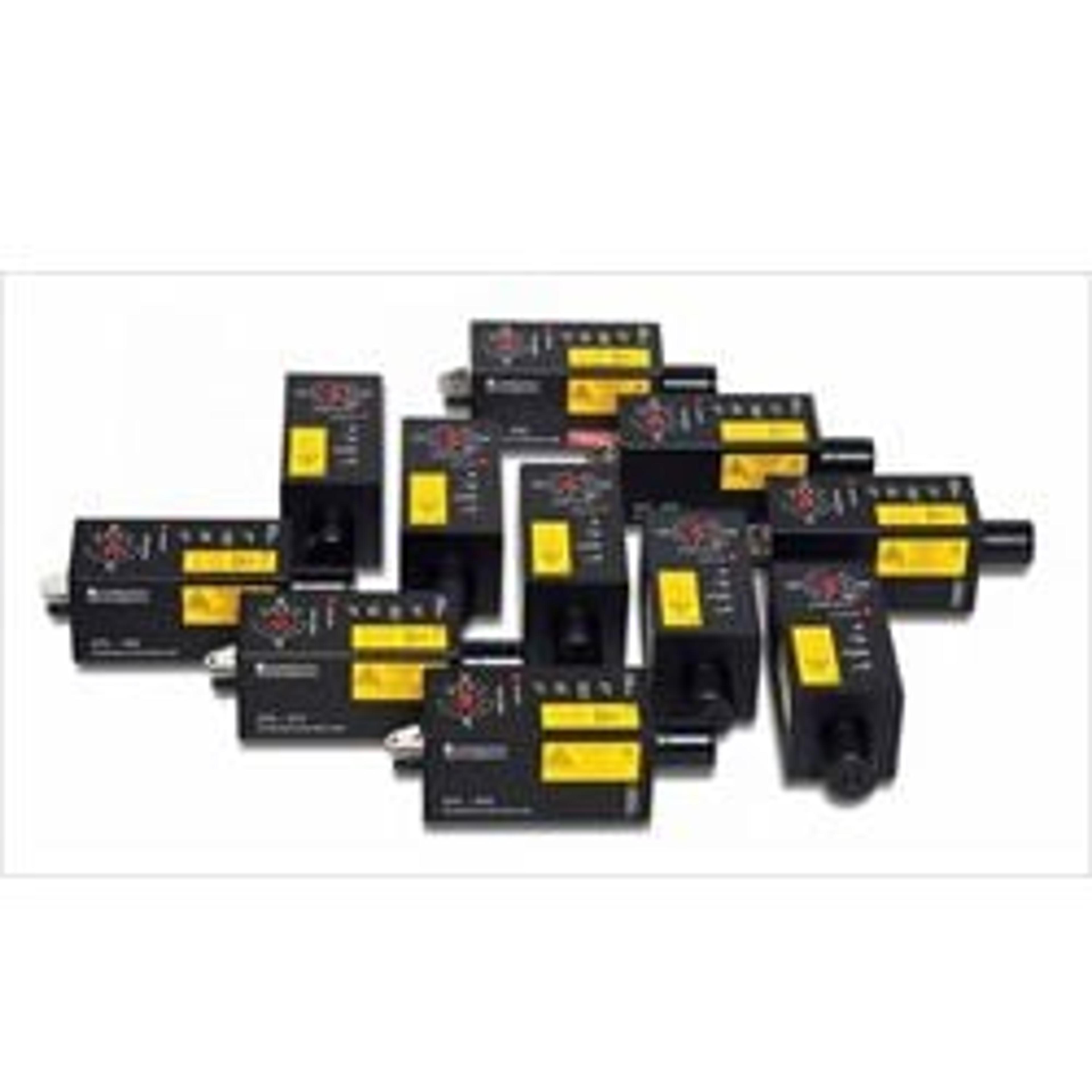
- Integrated Narrowband Raman Lasers: Accommodates up to three laser wavelengths, providing flexibility for application-specific optimization.
- High Optical Efficiency: Features free-space coupled lasers and a mirror-based spectrograph for optimal Raman signal acquisition.
- Software-Controlled Grating Turret: Supports up to five software-selectable gratings, ensuring optimal spectral resolution independent of the chosen laser.
- Integrated Detectors: Capable of integrating two detectors to cover a broad range of wavelengths and experimental requirements.
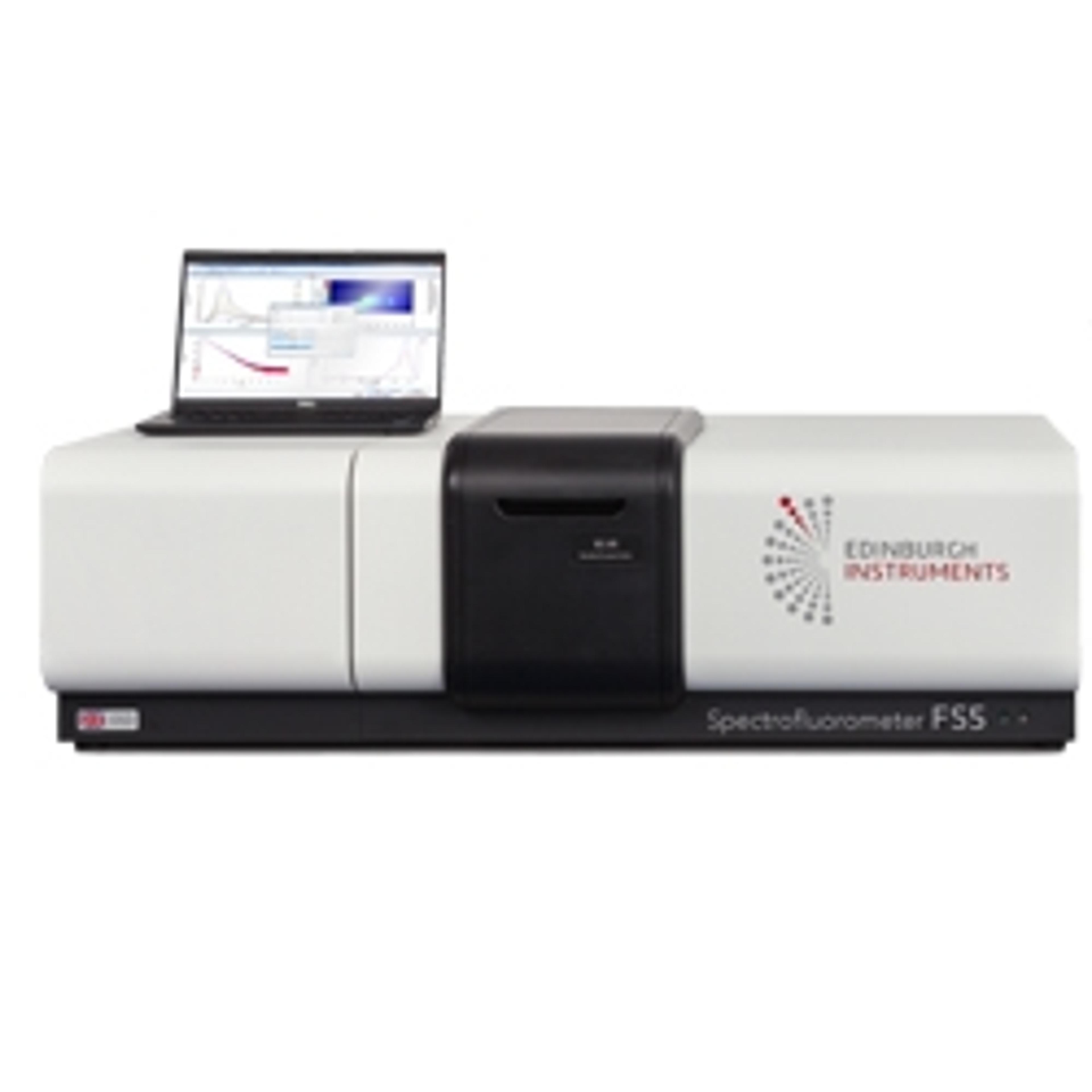
The RM5 features a streamlined yet comprehensive design, ensuring ease of use for Raman microscopy users of all experience levels. Ramacle® software guides users through all measurement steps while providing complete access to Raman capabilities, generating high-quality, publication-ready data.
Configurable with up to three fully software-controlled internal lasers spanning 405 nm to 1064 nm, the system maximises experimental flexibility within a compact footprint. Laser selection is complemented by a five-position grating turret, enabling the selection of gratings precisely matched to the chosen lasers. This integrated design optimises laboratory space and enhances user convenience by eliminating manual laser activation or grating exchange.
The RM5 enhances user experience with key functionalities: an internal silicon standard enables automated wavelength calibration, ensuring precise spectral measurements without user intervention. The confocal microscope offers an extensive range of pinhole positions for flexible measurement optimization. When coupled with a motorized stage, the software generates both 2D and 3D Raman and photoluminescence maps, along with SurfMAP® for analysing uneven surfaces.
This versatility enables the RM5 to integrate seamlessly into your laboratory, supporting a wide array of experiments across disciplines, making Raman microscopy accessible to biologists, chemists, and physicists alike.
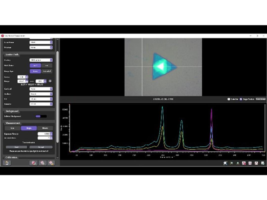
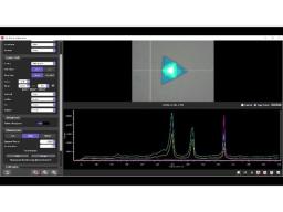
Spectral Measurements
Intuitive use is at the core of Ramacle’s design. We begin by focusing your sample using the microscope setup. Once you’re satisfied with the microscope image, we can proceed to the measurement setup window. The white-light image will carry over, allowing you to click on different areas of the sample for live Raman feedback using the live option. This feature helps you verify your measurement parameters and focus. You can then take single or multiple spectra from the selected sample area.
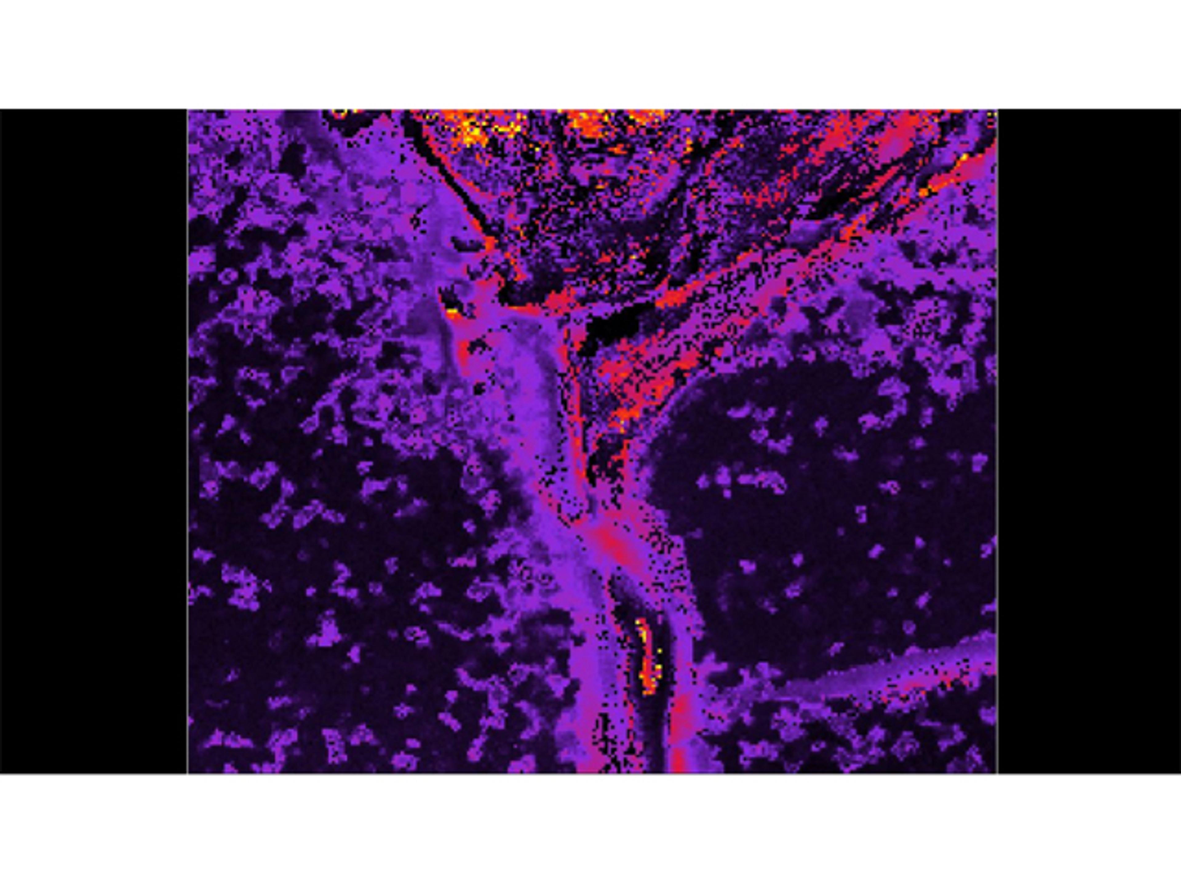
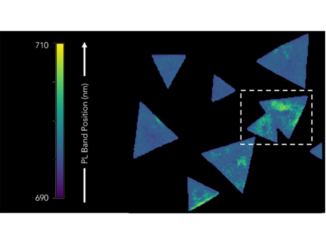
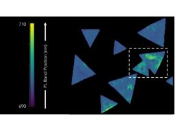
2D Mapping
2D mapping in Ramacle allows you to track the distribution of components in your sample. All the user needs to do is determine the area of interest to be investigated. Using Ramacle the area of interest to be mapped is defined, including areas larger than the field of view using stitched images. On the microscope image the map dimensions and step size are set for XY measurements, which will then move the stage to take a Raman spectrum from each point.
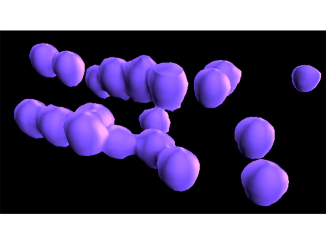
3D Mapping
Ramacle operates 3D mapping similarly to 2D mapping, with the added ability to define the map along the Z-axis. Thanks to the truly confocal nature of the RM5, highly resolved 3D maps can be captured. This advanced capability extends Raman mapping beyond surface analysis, constructing a detailed 3D chemical image at the micron scale.
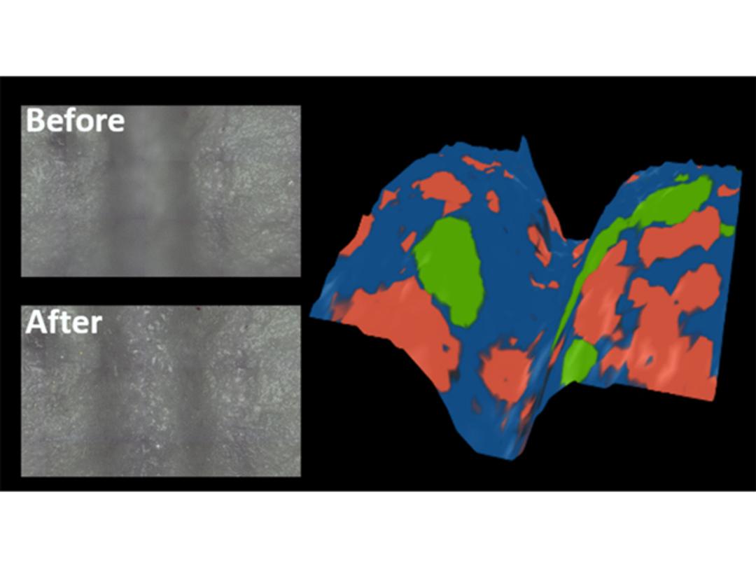
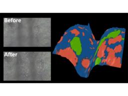
SurfMAP®
The SurfMAP® feature of Ramacle enables the user to analyse difficult samples with rough and uneven surfaces. This is done by adjusting the Z-axis to ensure the laser remains in perfect focus across the sample surface. Without this feature non-flat surfaces cannot be accurately mapped due to variation in signal caused by the laser moving out of focus to the sample.