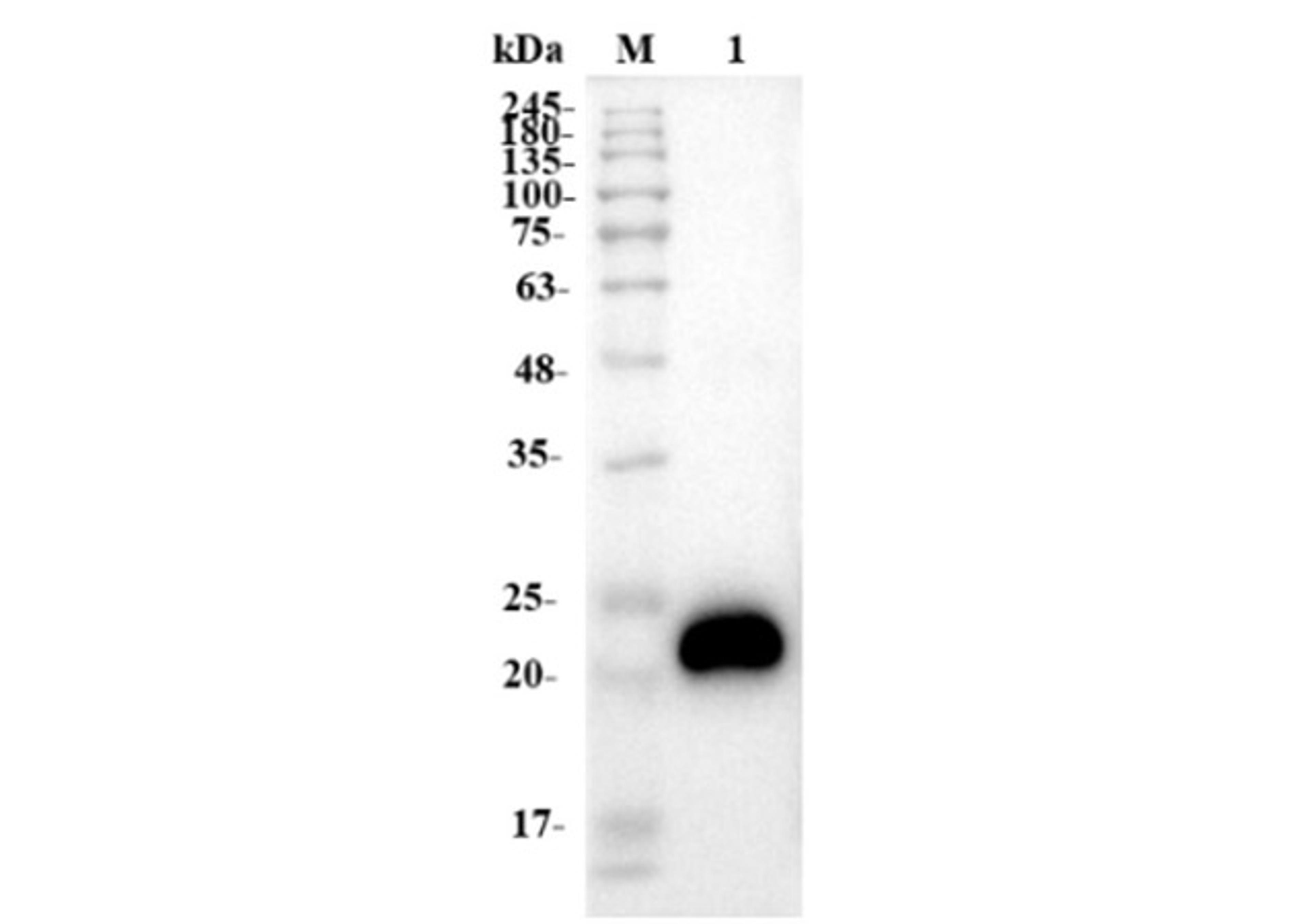
S100B
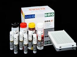
High Quality Assays with Reproducible and Reliable Results


The supplier does not provide quotations for this product through SelectScience. You can search for similar products in our Product Directory.
Immunoenzymatic colorimetric method for quantitative determination of S100B concentration in human serum and plasma.S100B ELISA kit is intended for laboratory use only.S100 is a 20 kDa protein belonging to the S100/calmodulin/troponin C superfamily of EF-hand calcium-binding proteins. S100 was originally isolated from human brain and considered a glial-cell specific protein (1). Today, 20 monomers of the S100 family have been identified based on structural and functional similarities (2,3). Most of the S100 proteins exist as dimers and are expressed in a cell-specific manner. Two of the S100 monomers, designated S100Alpha and S100Beta (4) are highly conserved between species and are found as homo- (BetaBeta) and heterodimers (AlphaBeta) in central nervous system glial cells and in certain peripheral cells e.g. Schwann cells, melanocytes, adipocytes, and chondrocytes (5). S100AlphaBeta and S100BetaBeta are also present in malignant tissues, most notably in melanoma and to a lesser extent in glioma, thyroid cell carcinoma and renal cell carcinoma (2). Determinationof S100B (like S100AlphaBeta and S100BetaBeta units) in serum has been shown to be clinically useful for prognosis and treatment monitoring of patients diagnosed with malignant melanoma (6-9). Studies also suggest that S100B may be useful in the management of patients with brain damage from e.g. traumatic head injury, perinatal asphyxia, cardiac arrest, cardiac surgery and stroke (10-13).The S100B ELISA TEST is based on binding of S100B by two antibodies, one immobilized on microwell plates, and the other one conjugates with horseradish peroxidase (HRP). The assay is a two steps binding procedure and after every incubation step, the bound/free separation is performed by a simple solid-phase washing. Then, the enzyme HRP in the bound-fraction reacts with the Substrate (H2O2) and the TMB Substrate and develops a blue color that changes into yellow when the Stop Solution (H2SO4) is added. The color intensity is proportional to the S100B concentration in the sample. S100B concentration in the sample is calculated through a calibration curve.