sVCAM-1 human
High Quality Assays with Reproducible and Reliable Results

The supplier does not provide quotations for this product through SelectScience. You can search for similar products in our Product Directory.
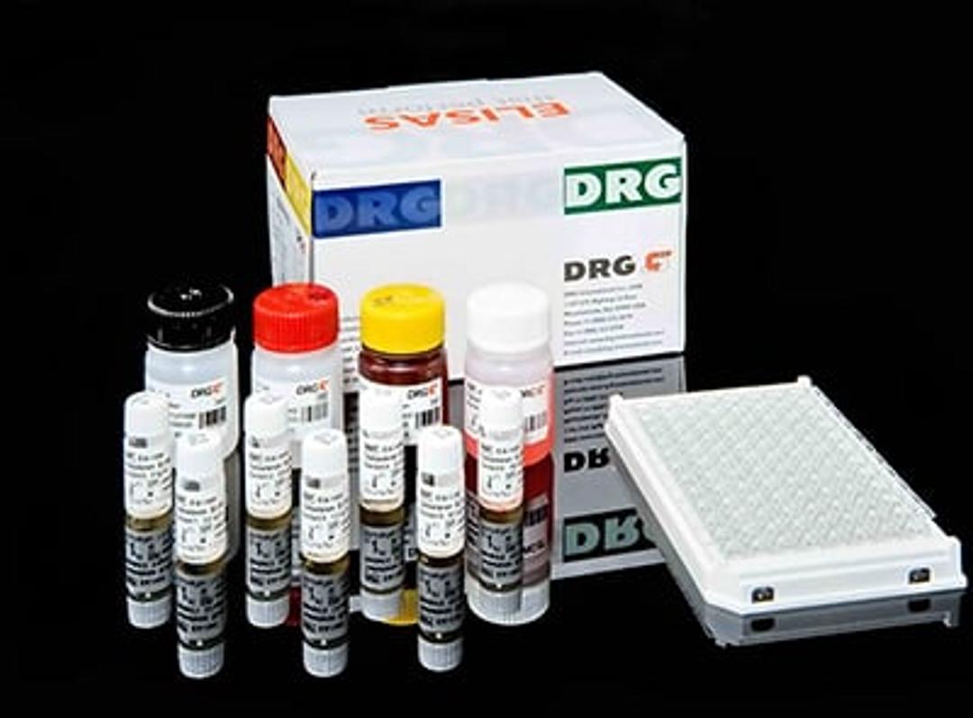
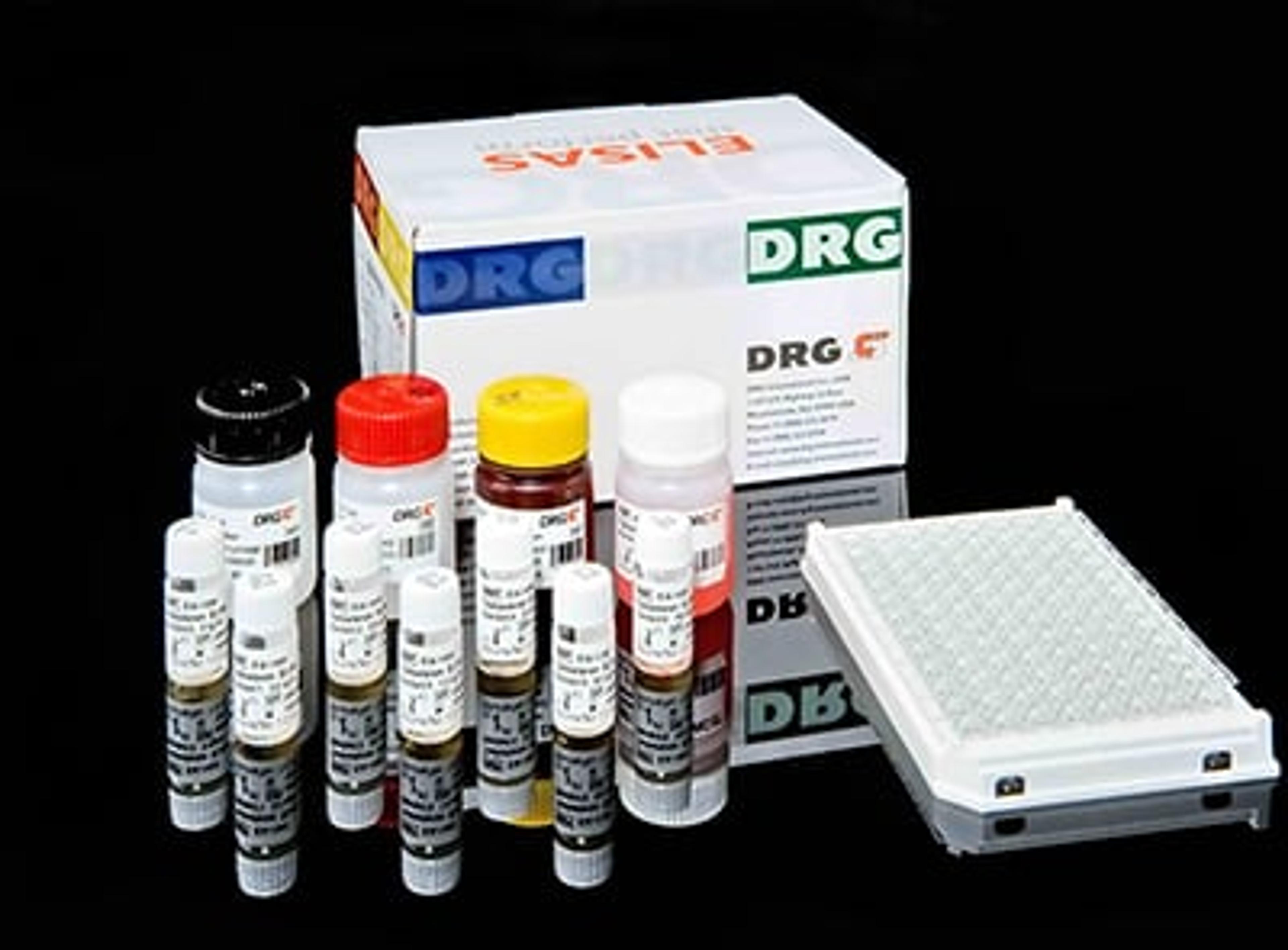
The human sVCAM-1 ELISA is an enzyme-linked immunosorbent assay for the quantitative detection of human sVCAM-1.The human sVCAM-1 ELISA is for in vitro diagnostic use. Not for use in therapeutic procedures.The vascular cell adhesion molecule-1 (VCAM-1) or CD106 is a member of the immunoglobulin gene superfamily. The initial molecular cloning of VCAM-1 reported six extracellular Ig-like domains (6D VCAM-1). This 6D VCAM-1 arises due to alternative splicing from a seven-domain VCAM-1 (7D VCAM-1). 7D VCAM-1 is the dominant form expressed by cultured human endothelial cells. Domains 1 through 3 are highly homologous to domains 4 through 6, suggesting that they arose by gene duplication. The cDNA of 7D VCAM-1 predicts a core protein of approximately 81 kDa with seven potential N-linked glycosylation sites. Upon complete glycosylation the mature protein has a molecular weight of approximately 102 kDa. This observation is in general agreement with immunoprecipitation studies that show a protein of approximately 110 kDa on cytokine-activated endothelium. Murine and rat VCAM-1 have been cloned. In contrast to ICAM-1, VCAM-1 appears to have been highly conserved through evolution. Both rat and mouse VCAM-1 are highly homologous at the protein level to the human VCAM-1 (77% and 76%, respectively). VCAM-1 supports the adhesion of lymphocytes, monocytes, natural killer cells,eosinophils, and basophils through its interaction with leukocyte very late antigen-4 (VLA-4). VCAM-1/VLA-4 interaction mediates firm adherence of circulating non-neutrophilic leukocytes to endothelium. VCAM-1 also participates in leukocyte adhesion outside of the vasculature, mediating precursor lymphocyte adhesion to bone marrow stromal cells and B cell binding to lymph node follicular dendritic cells. VCAM-1 is not constitutively expressed on endothelium, but can be up-regulated in vitro in response to LPS, TNF-a, andIL-1, as well as to interferon-g and IL-4. VCAM-1 is also present on tissue macrophages, dendritic cells, bone marrow fibroblasts, myoblasts and myotubes. A soluble form of VCAM-1 (sVCAM-1) has been described. Soluble VCAM-1 levels have been found in the serum of healthy individuals and increased levels of sVCAM-1 can be detected in several diseases:Cancer: ovarian, gastric-intestinal, renal, bladder cancer, non-Hodgkin's lymphoma, breast cancer (cyst fluid);Autoimmune diseases: multiple sclerosis (cerebrospinal fluid), systemic sclerosis, systemic lupus erythematosus, rheumatoid arthritis;Infections: sepsis, meningitis, malaria;Inflammation: vasculitis, alcoholic cirrhosis, primary biliary cirrhosis, Wegener's granulomatosis;Others: impaired renal function,haemodialysis, hyperthyroidism, renal allograft. Ananti-human sVCAM-1 coating antibody is adsorbed ontomicrowells.Human sVCAM-1present in the sample or standard binds to antibodies adsorbed to themicrowells. A conjugate mixture (biotin-conjugated anti-humansVCAM-1 antibody and Streptavidin-HRP) is added. Biotin-conjugatedanti-human sVCAM-1 antibody binds to human sVCAM-1captured by the first antibody.Streptavidin-HRPbinds to the biotin-conjugated anti-human sVCAM-1antibody.Followingincubation unbound Streptavidin-HRP is removed during a wash step, andsubstrate solution reactive with HRP is added to the wells.A coloured product is formed in proportion to theamount of human sVCAM-1 present in the sample orstandard. The reaction is terminated by addition of acid and absorbance ismeasured at 450 nm. A standard curve is prepared from 6 humansVCAM-1 standard dilutions and human sVCAM-1sample concentration determined.

