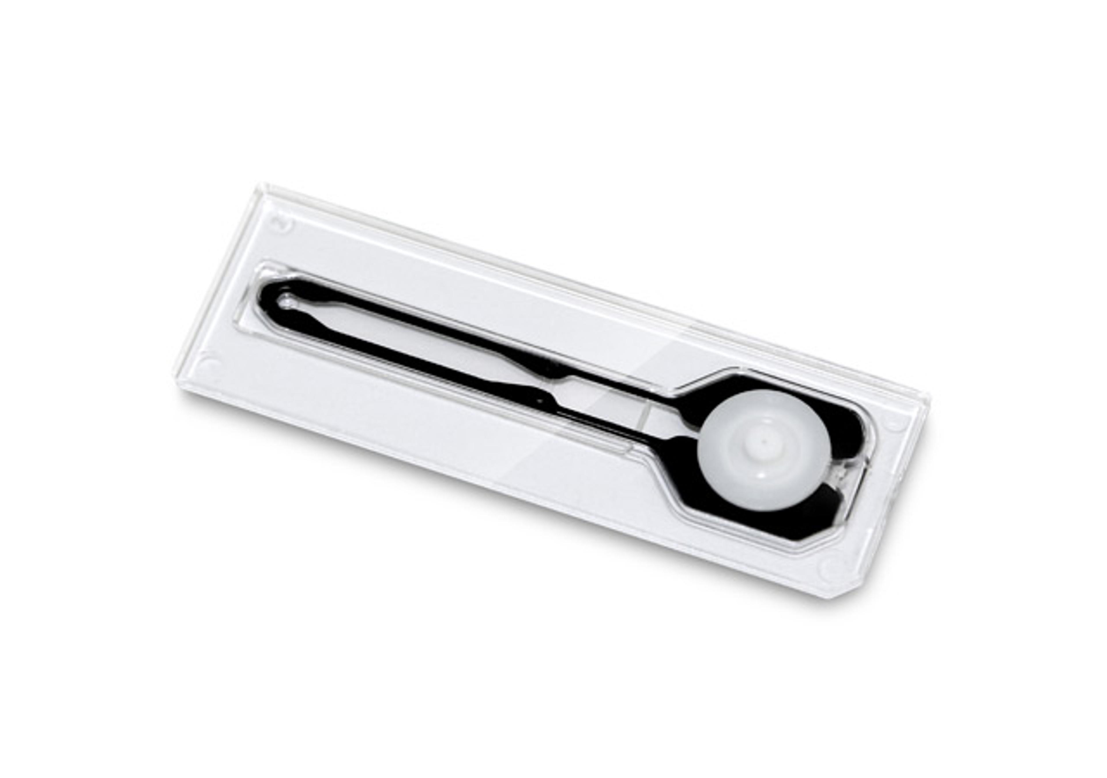
TPO (Thyroid Peroxidase)
High Quality Assays with Reproducible and Reliable Results

The supplier does not provide quotations for this product through SelectScience. You can search for similar products in our Product Directory.
The Quantitative Determination of Thyroid Peroxidase (TPO) Autoantibodies in Human Serum or Plasma by a Microplate Enzyme Immunoassay. Measurements of TPO autoantibodies may aid in the diagnosis of certain thyroid diseases such as Hashimoto’s and Grave’s as well as nontoxic goiter.Antibodies to thyroid peroxidase have been shown to be characteristically present from patients with Hashimoto thyroiditis (95%), idiopathic myedema (90%) and Graves Disease (80%)1. In fact 72% of patients positive for anti-TPO exhibit some degree of thyroid dysfunction. This has led to the clinical measurement becoming a valuable tool in the diagnosis of thyroid dysfunction. Measurements of antibodies to TPO have been done, in the past, by Passive Hemaglutination (PHA). PHA tests do not have the sensitivity of enzyme immunoassay and are limited by subjective interpretation. This procedure, with the enhanced sensitivity of EIA, permits the detectability of subclinical levels of antibodies to TPO. In addition, the results are quantitated by a spectrophotometer, which eliminates subjective interpretation. The microplate enzyme immunoassay methodology provides the technician with optimum sensitivity while requiring few technical manipulations. In this method, serum reference, diluted patient specimen, or control is first added to a microplate well. Biotinylated Thyroid Peroxidase Antigen (TPO) is added, and then the reactants are mixed. Reaction results between the autoantibodies to TPO and the biotinylated TPO toform an immune complex, which is deposited to the surface of streptavidin coated wells through the high affinity reaction of biotin and streptavidin. After the completion of the required incubation period, aspiration or decantation separates the reactants that are not attached to the wells. An enzyme anti-human IgG conjugate is then added to permit quantitation of reaction through interacting with human IgG of the immune complex. After washing, the enzyme activity is determined by reaction with substrate to produce color.The employment of several serum references of known antibody activity permits construction of a graph of enzyme and antibody activities. From comparison to the dose response curve, an unknown specimen’s enzyme activity can be correlated with autoimmune antibody level.

